Abstract
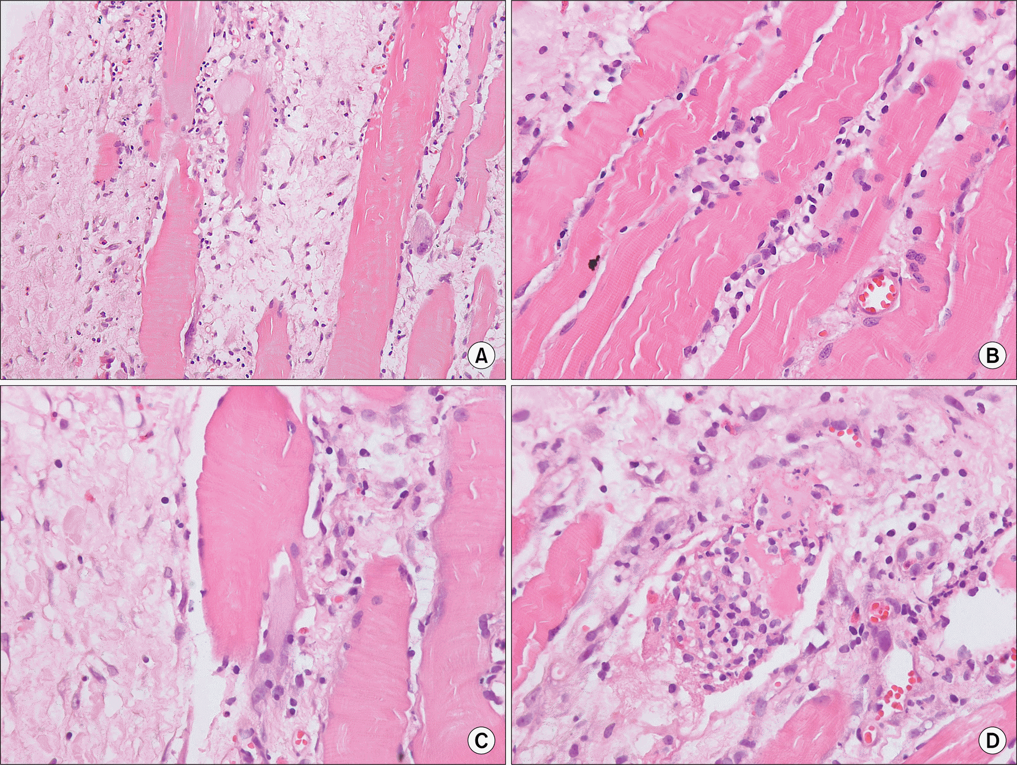
Exercise-induced muscle damage (EIMD) can be caused by novel or unaccustomed exercise resulting in a temporary decrease in muscle function, increased muscle soreness and swelling, and an increase in muscle proteins in blood. A 38-year-old female presented with a 2-week history of bilateral lower leg pain and swelling that developed suddenly after performing 108 prostrations. Fat-suppressed contrastenhanced T1-weighted magnetic resonance imaging showed bilateral symmetric high signal intensity of the tibialis anterior muscles. Our patient was diagnosed with acute myositis and treated with naproxen. History of physical exertion and acute onset of typical clinical findings of myositis were key elements in the diagnosis. Acute myositis related to exercise is a common and self-limiting condition which fully resolves after 2 to 3 weeks. However, other causes of myositis should be excluded in patients with atypical clinical features.
REFERENCES
1. Brentano MA, Martins Kruel LF. A review on strength exercise-induced muscle damage: applications, adaptation mechanisms and limitations. J Sports Med Phys Fitness. 2011; 51:1–10.
2. Hicks KM, Onambélé GL, Winwood K, Morse CI. Muscle damage following maximal eccentric knee extensions in males and females. PLoS One. 2016; 11:e0150848.

3. Lieber RL, Fridén J. Muscle damage is not a function of muscle force but active muscle strain. J Appl Physiol (1985). 1993; 74:520–6.

4. Malm C. Exercise-induced muscle damage and inflammation: fact or fiction? Acta Physiol Scand. 2001; 171:233–9.

5. Neubauer O, König D, Wagner KH. Recovery after an Ironman triathlon: sustained inflammatory responses and muscular stress. Eur J Appl Physiol. 2008; 104:417–26.

6. Clarkson PM, Hubal MJ. Exercise-induced muscle damage in humans. Am J Phys Med Rehabil. 2002; 81(11 Suppl):S52–69.

7. MacIntyre DL, Reid WD, McKenzie DC. Delayed muscle soreness. The inflammatory response to muscle injury and its clinical implications. Sports Med. 1995; 20:24–40.
8. Clarkson PM, Nosaka K, Braun B. Muscle function after exercise-induced muscle damage and rapid adaptation. Med Sci Sports Exerc. 1992; 24:512–20.

9. Schulze M, Kötter I, Ernemann U, Fenchel M, Tzaribatchev N, Claussen CD, et al. MRI findings in inflammatory muscle diseases and their noninflammatory mimics. AJR Am J Roentgenol. 2009; 192:1708–16.

10. Nosaka K, Clarkson PM. Changes in indicators of inflammation after eccentric exercise of the elbow flexors. Med Sci Sports Exerc. 1996; 28:953–61.

Figure 1.
Magnetic resonance imaging of both lower legs. T1-weighted images (A, coronal and B, axial view) showing iso-signal intensity (arrows). Fat-suppressed contrastenhanced T1-weighted images (C, D) showing bilateral symmetric high signal intensity of the tibialis anterior muscles and of the anterior portion fascia (arrowheads).


 PDF
PDF ePub
ePub Citation
 Print
Print


 XML Download