Journal List > J Rheum Dis > v.23(6) > 1064291

Lim, Kwon, Jung, and Park: Tumor Necrosis Factor Blockade Stimulates Circulating Osteoblastic Lineage Cells Activity while Reducing Circulating Osteoclasts

Abstract

Objective

This study examines the effects of tumor necrosis factor (TNF) blockade on markers of bone metabolism in peripheral blood from active rheumatoid arthritis (RA) patients.

Methods

Eighteen patients (16 women, 2 men) aged 50 years (range 37-63 years), with persistently active RA (mean disease duration 7 years) were studied. Most took methotrexate (mean dose 12.5 mg) and all except one received corticosteroid (mean dose 5.7 mg). Four were treated with etanercept, eight received adalimumab and six received infliximab. Before and six months after taking TNF blockers, blood was sampled to obtain peripheral blood mononuclear cells (PBMCs), and serum bone turnover markers and acute phase reactants were measured. PBMCs were seeded and cultured to produce osteoblastic lineage cells and osteoclasts.

Results

The formation of calcified nodules by osteoblastic lineage cells from PBMC increased from 205.7±196.3 μ mol/well at the baseline to 752.5±671.9 μ mol/well after TNF blockade (p<0.024). The serum levels of bone formation markers, including bone specific alkaline phosphatase and osteocalcin also increased. The number of circulating osteoclasts and area of bone resorption pits made by osteoclasts were reduced after TNF blockade.

Conclusion

The activity of circulating osteoblastic lineage cells increased after TNF blockade, whereas peripheral osteoclastogenesis tended to be suppressed. This is the first study of cultured human peripheral osteoblastic lineage cells in RA patients. Given that peripheral bone formation is difficult to study using radiologic methods, culture of these cells may provide a new modality for studying bone metabolism in RA.

REFERENCES

1. Kotake S, Nanke Y. Effect of TNFα on osteoblastogenesis from mesenchymal stem cells. Biochim Biophys Acta. 2014; 1840; 1209–13.
crossref
2. McInnes IB, Schett G. The pathogenesis of rheumatoid arthritis. N Engl J Med. 2011; 365:2205–19.
crossref
3. Breedveld FC, Weisman MH, Kavanaugh AF, Cohen SB, Pavelka K, van Vollenhoven R, et al. The PREMIER study: a multicenter, randomized, double-blind clinical trial of combination therapy with adalimumab plus methotrexate versus methotrexate alone or adalimumab alone in patients with early, aggressive rheumatoid arthritis who had not had previous methotrexate treatment. Arthritis Rheum. 2006; 54:26–37.
crossref
4. Emery P, Breedveld FC, Hall S, Durez P, Chang DJ, Robertson D, et al. Comparison of methotrexate monotherapy with a combination of methotrexate and etanercept in active, early, moderate to severe rheumatoid arthritis (COMET): a randomised, double-blind, parallel treatment trial. Lancet. 2008; 372:375–82.
crossref
5. Szentpetery A, McKenna MJ, Murray BF, Ng CT, Brady JJ, Morrin M, et al. Periarticular bone gain at proximal interphalangeal joints and changes in bone turnover markers in response to tumor necrosis factor inhibitors in rheumatoid and psoriatic arthritis. J Rheumatol. 2013; 40:653–62.
crossref
6. Finzel S, Rech J, Schmidt S, Engelke K, Englbrecht M, Stach C, et al. Repair of bone erosions in rheumatoid arthritis treated with tumour necrosis factor inhibitors is based on bone apposition at the base of the erosion. Ann Rheum Dis. 2011; 70:1587–93.
crossref
7. Døhn UM, Ejbjerg B, Boonen A, Hetland ML, Hansen MS, Knudsen LS, et al. No overall progression and occasional repair of erosions despite persistent inflammation in adalimumab-treated rheumatoid arthritis patients: results from a longitudinal comparative MRI, ultrasonography, CT and radiography study. Ann Rheum Dis. 2011; 70:252–8.
8. Prevoo ML, van't Hof MA, Kuper HH, van Leeuwen MA, van de Putte LB, van Riel PL. Modified disease activity scores that include twenty-eight-joint counts. Development and validation in a prospective longitudinal study of patients with rheumatoid arthritis. Arthritis Rheum. 1995; 38:44–8.
crossref
9. Lange U, Teichmann J, Müller-Ladner U, Strunk J. Increase in bone mineral density of patients with rheumatoid arthritis treated with anti-TNF-alpha antibody: a prospective open-label pilot study. Rheumatology (Oxford). 2005; 44:1546–8.
10. Dischereit G, Tarner IH, Müller-Ladner U, Lange U. Infliximab improves bone metabolism and bone mineral density in rheumatoid arthritis and ankylosing spondylitis: a prospective 2-year study. Clin Rheumatol. 2013; 32:377–81.
crossref
11. Kuznetsov SA, Mankani MH, Gronthos S, Satomura K, Bianco P, Robey PG. Circulating skeletal stem cells. J Cell Biol. 2001; 153:1133–40.
crossref
12. Friedenstein AJ. Osteogenic stem cells in bone marrow. Heersche JNM, Kanis JA, editors. Bone and mineral research. Amsterdam: Elsevier;1990. p. 243–72.
13. Pittenger MF, Mackay AM, Beck SC, Jaiswal RK, Douglas R, Mosca JD, et al. Multilineage potential of adult human mesenchymal stem cells. Science. 1999; 284:143–7.
crossref
14. Long MW, Williams JL, Mann KG. Expression of human bone-related proteins in the hematopoietic microenvironment. J Clin Invest. 1990; 86:1387–95.
crossref
15. Long MW, Robinson JA, Ashcraft EA, Mann KG. Regulation of human bone marrow-derived osteoprogenitor cells by osteogenic growth factors. J Clin Invest. 1995; 95:881–7.
crossref
16. Eghbali-Fatourechi GZ, Lamsam J, Fraser D, Nagel D, Riggs BL, Khosla S. Circulating osteoblast-lineage cells in humans. N Engl J Med. 2005; 352:1959–66.
crossref
17. Kumagai K, Vasanji A, Drazba JA, Butler RS, Muschler GF. Circulating cells with osteogenic potential are physiologically mobilized into the fracture healing site in the para-biotic mice model. J Orthop Res. 2008; 26:165–75.
crossref
18. Matsuno H, Yoshida K, Ochiai A, Okamoto M. Requirement of methotrexate in combination with anti-tumor necrosis factor-alpha therapy for adequate suppression of osteoclastogenesis in rheumatoid arthritis. J Rheumatol. 2007; 34:2326–33.
19. Gengenbacher M, Sebald HJ, Villiger PM, Hofstetter W, Seitz M. Infliximab inhibits bone resorption by circulating osteoclast precursor cells in patients with rheumatoid arthritis and ankylosing spondylitis. Ann Rheum Dis. 2008; 67:620–4.
crossref
20. Dalle Carbonare L, Valenti MT, Zanatta M, Donatelli L, Lo Cascio V. Circulating mesenchymal stem cells with abnormal osteogenic differentiation in patients with osteoporosis. Arthritis Rheum. 2009; 60:3356–65.
crossref

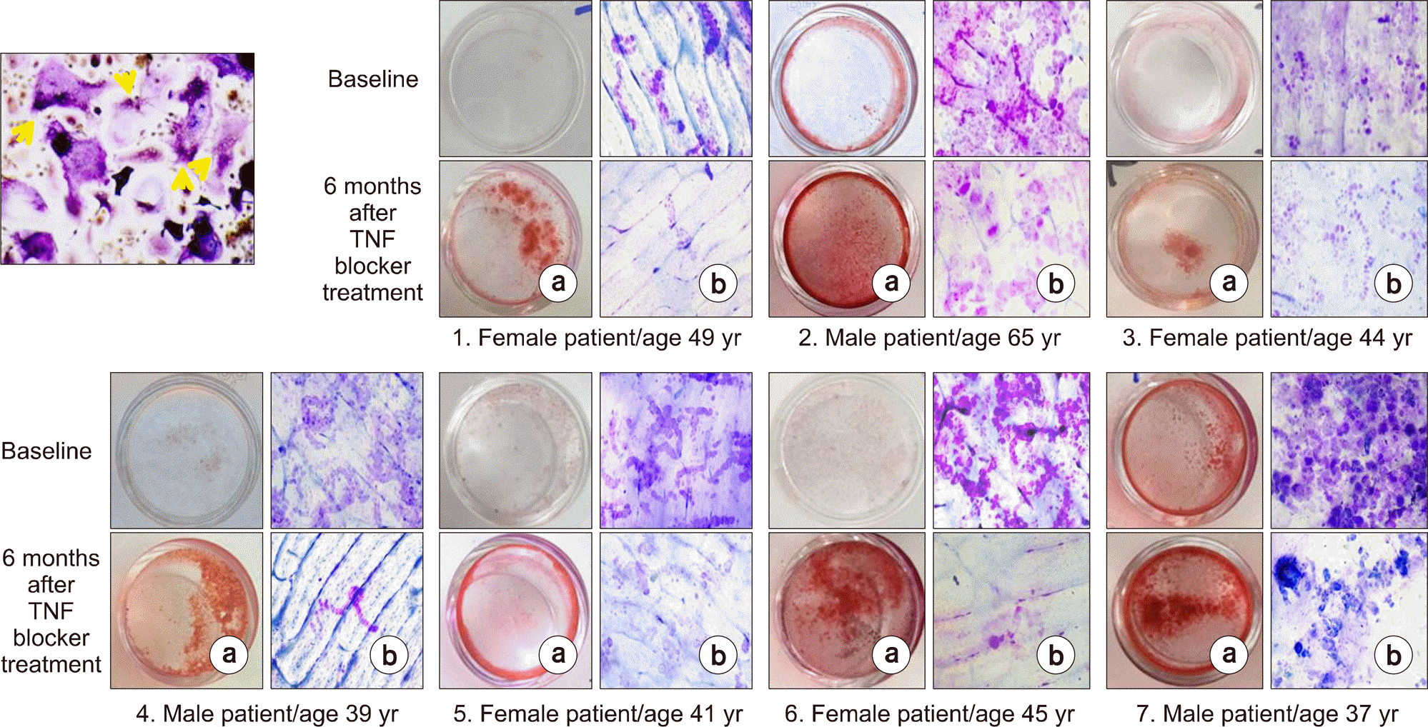
Figure 1.
Calcified nodule formation (a) and bone resorption pits (b) before and after tumor necrosis factor (TNF) blocker treatment. The multinucleated osteoclasts which are responsible for bone resorption is shown in left upper corner. Patients 1∼3 were treated with adalimumab, patients 4∼5 with etanercept and patients 6∼7 with infliximab. TRAP staining, 200×.
jrd-23-356f1.tif
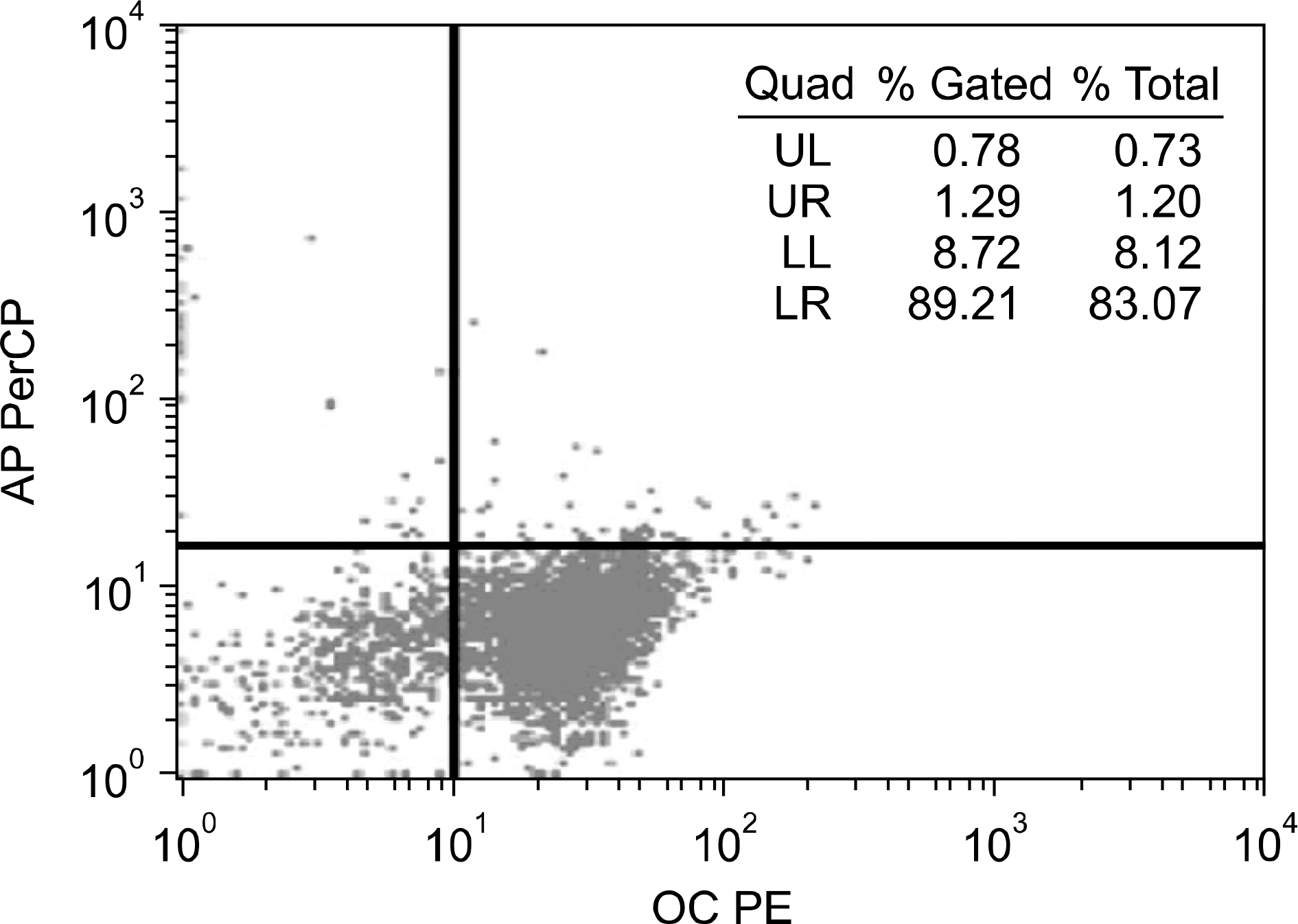
Figure 2.
Flow cytometry analysis of cells from peripheral blood to sort for both osteocalcin, bone specific alkaline phosphatase (BSALP)-positive cells. Cells positive for osteocalcin (OC) were stained with phycoerythrin (PE) and cells positive for BSALP (AP) were conjugated with peridinin chlorophyll protein (PerCP). Cells positive for both osteocalcin and BSALP were turned out to be less than 1%. UL: upper left, UR: upper right, LL: lower left, LR: lower right, Quad: quadrant.
jrd-23-356f2.tif
Table 1.
Demographics of patients enrolled in the study
Variable Patient (n=18)
TNF blockers used (n)  
 Etanercept 4
 Adalimumab 8
 Infliximab 6
 Age (yr) 49.7±8.3
 Women (%) 88.9
 Disease duration (yr) 7.0±6.6
 Dose of prednisolone (mg/d) 5.7±2.7
 Duration of prednisolone use (yr) 5.6±6.4
 Methotrexate prescribed (%) 94.4
 Dose of methotrexate (mg/wk) 12.5±4.46
 Calcium and Vitamin D prescribed (%) 50.5
 Bisphosphonates prescribed (%) 0

Values are number only or mean±standard deviation. TNF: tumor necrosis factor.

Table 2.
Changes in disease activities and biochemical markers of bone turnover after TNF blocker treatment
Variable At baseline At 6 months p-value
Tender joint counts 16±7 8.0±5.7 <0.001
Swollen joint counts 16.0±7.3 8.1±5.6 0.001
Pain scale 75.0±14.2 40.0±23.8 <0.001
ESR (mm/hr) 34.0±19.6 28.0±28.1 0.148
CRP (mg/dL) 1.64±1.44 0.86±1.68 0.124
DAS28-ESR 6.62±0.93 4.75±1.57 <0.001
DAS28-CRP 6.2±0.8 4.24±1.31 <0.001
IL-6 (pg/mL) 23.82±26.40 17.06±32.50 0.54
BSALP (U/L) 22.17±6.99 25.82±8.89 0.007
Osteocalcin (ng/mL) 5.81±1.94 8.60±3.23 <0.001
CTX (ng/mL) 0.52±0.27 0.64±0.29 0.118
Cathepsin K (pmol/L) 4.99±1.97 4.17±2.08 0.123

Values are presented as mean±standard deviation. DAS28 is Disease Activity Score based on 28 joints for rheumatoid arthritis patients [8]. TNF: tumor necrosis factor, ESR: erythrocyte sedimentation rate, CRP: C-reactive protein, IL: interleukin, BSALP: bone specific alkaline phosphatase, CTX: c-terminal telopeptide.

Table 3.
Changes in disease activities and biochemical markers of bone turnover in DAS28 responders after TNF blocker treatment
Variable At baseline At 6 months p-value
ESR (mm/hr) 29.0±17.7 17.0±14.3 0.002
CRP (mg/dL) 1.60±1.42 0.20±0.47 0.001
DAS28-ESR 6.48±0.94 4.09±0.96 <0.001
DAS28-CRP 6.19±0.81 3.68±0.68 <0.001
IL-6 (pg/mL) 28.07±28.32 10.52±29.72 0.149
BSALP (U/L) 22.48±7.62 26.07±9.83 0.023
Osteocalcin (ng/mL) 5.87±1.61 8.59±2.86 <0.001
CTX (ng/mL) 0.48±0.19 0.66±0.31 0.018
Cathepsin K (pmol/L) 4.66±1.80 3.69±2.26 0.1

Values are presented as mean±standard deviation. DAS28 is Disease Activity Score based on 28 joints for rheumatoid arthritis patients [8]. TNF: tumor necrosis factor, ESR: erythrocyte sedimentation rate, CRP: C-reactive protein, IL: interleukin, BSALP: bone specific alkaline phosphatase, CTX: c-terminal telopeptide.

Table 4.
Changes in optical densities of osteoblasts, number of osteoclasts and area of bone resorption pits in addition to disease activities and biochemical markers of bone turnover in DAS28 non-responders after TNF blocker treatment
Variable At baseline At 6 months p-value
Optical density of calcified nodule (μ mol/well) 133.90±184.77 202.8±266.84 0.784
Bone resorption pit by osteoclasts (%) 29.0±28.4 45.0±3.4 0.392
Number of osteoclasts (/well) 547.0±419.1 437.0±124.8 0.655
ESR (mm/hr) 53.0±15.8 67.0±30.9 0.465
CRP (mg/dL) 1.80±1.72 3.10±2.56 0.465
DAS28-ESR 7.10±0.82 7.00±1.03 0.715
DAS28-CRP 6.3±0.9 6.18±1.09 1.0
IL-6 (pg/mL) 8.97±9.90 39.97±35.72 0.465
BSALP (U/L) 20.70±3.16 24.70±1.53 0.162
Osteocalcin (ng/mL) 5.52±3.62 8.66±5.51 0.102
CTX (ng/mL) 0.72±0.52 0.58±0.14 1.0
Cathepsin K (pmol/L) 5.92±2.42 5.47±0.43 0.715

Values are presented as mean±standard deviation. DAS28 is Disease Activity Score based on 28 joints for rheumatoid arthritis patients [8]. TNF: tumor necrosis factor, ESR: erythrocyte sedimentation rate, CRP: C-reactive protein, IL: interleukin, BSALP: bone specific alkaline phosphatase, CTX: c-terminal telopeptide.

TOOLS
Similar articles