Abstract
Intraarticular hyaluronic acid injections for symptomatic treatment of osteoarthritis are widely used but can result in complications, such as infectious arthritis. Staphylococcus lugdunensis is a common normal skin flora but can cause severe infectious disease, such as infective endocarditis. We present the first report of infectious arthritis caused by methicillin-sensitive S. lugdunensis after intra-articular hyaluronic acid injection in an immunocompromised patient in Korea.
REFERENCES
1. Frank KL, Reichert EJ, Piper KE, Patel R. In vitro effects of antimicrobial agents on planktonic and biofilm forms of Staphylococcus lugdunensis clinical isolates. Antimicrob Agents Chemother. 2007; 51:888–95.
2. Bernardeau C, Bucki B, Lioté F. Acute arthritis after intraarticular hyaluronate injection: onset of effusions without crystal. Ann Rheum Dis. 2001; 60:518–20.

3. Shemesh S, Heller S, Salai M, Velkes S. Septic arthritis of the knee following intraarticular injections in elderly patients: report of six patients. Isr Med Assoc J. 2011; 13:757–60.
4. Freney J, Brun Y, Bes M, Meugnier H, Grimont F, Grimond PAD, et al. Staphylococcus lugdunensis sp. Nov. and Staphylococcus schleiferi sp. Nov., two species from human clinical specimens. Int J Syst Evol Microbiol. 1988; 38:168–72.

5. Maria M, Manuel R, Clara C, Juan G. Artritis séptica por Staphylococcus lugdunensis. Reumatol Clin. 2009; 5:44–5.
6. Klotchko A, Wallace MR, Licitra C, Sieger B. Staphylococcus lugdunensis: an emerging pathogen. South Med J. 2011; 104:509–14.
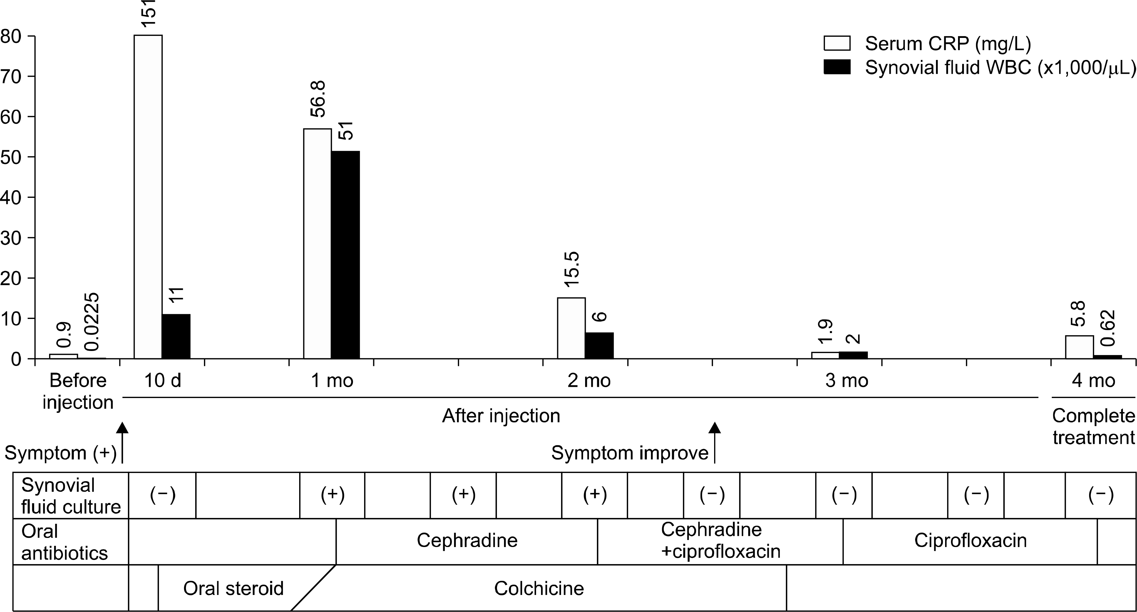
Figure 1.
The patient's clinical course was improved after antibiotics treatment. WBC: white blood cell, CRP: C-reactive protein, d: day, mo: month.

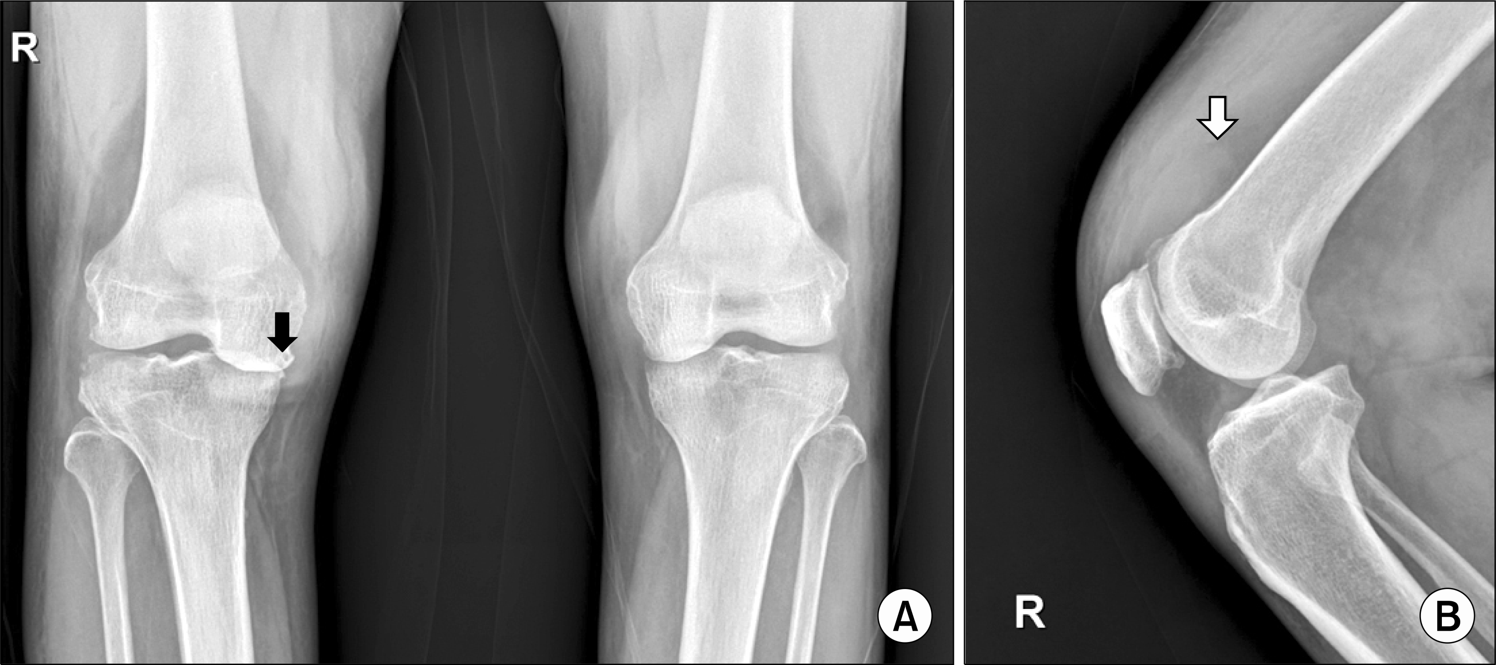
Figure 2.
(A) The anteroposterior view of both knee. (B) The lateral view of right knee. Marginal spurs and mild subchondral sclerosis in medial femo-rotibial (black arrow) & patellofemoral compartments at right knee joint. Marginal erosion and juxtaarticular osteoporosis at lateral tibial plateau are noted in the right knee joint with large amount effusion (white arrow).


 PDF
PDF ePub
ePub Citation
 Print
Print


 XML Download