

 PDF
PDF ePub
ePub Citation
 Print
Print


Abstract
Rice bodies are materials with an amorphous nucleus and a fibrin layer found floating in the synovial space and bursa. These bodies have often been detected in patients with rheumatoid arthritis, tuberculous arthritis, and bursitis. Although the etiology and pathogenesis of rice bodies are not yet fully understood, it has been hypothesized that they might be caused by chronic inflammation originating from the synovium. However, we report on a case of idiopathic massive rice bodies in the knee joint without evidence of inflammatory articular disease or infection including rheumatoid arthritis, seronegative spondyloarthritides, tuberculosis, or bacterial or fungal infection.
Go to : 
REFERENCES
1. Reise H. Die Reiskörperchen in tuberculös erkrankten Synovialsäcken. Deutsch Z Chir. 1895; 42:1–99.
2. Popert AJ, Scott DL, Wainwright AC, Walton KW, Williamson N, Chapman JH. Frequency of occurrence, mode of development, and significance or rice bodies in rheumatoid joints. Ann Rheum Dis. 1982; 41:109–17.

3. Thevenon A, Cocheteux P, Duquesnoy B, Mestdagh H, Lecomte-Houcke M, Delcambre B. Subacromial bursitis with rice bodies as a presenting feature of seronegative rheumatoid arthritis. Arthritis Rheum. 1987; 30:715–6.
4. Bulut M, Yilmaz E, Karakurt L, Özercan MR. Rice body formation characterized by the chronic non-specific tenosynovitis in the tibialis anterior tendon. Acta Orthop Traumatol Turc. 2013; 47:142–5.
5. Kim RS, Lee JY, Jung SR, Lee KY. Tuberculous subdeltoid bursitis with rice bodies. Yonsei Med J. 2002; 43:539–42.
6. Chau CL, Griffith JF, Chan PT, Lui TH, Yu KS, Ngai WK. Rice-body formation in atypical mycobacterial tenosynovitis and bursitis: findings on sonography and MR imaging. AJR Am J Roentgenol. 2003; 180:1455–9.
7. Kassimos D, George E, Kirwan JR. Rice bodies in the pleural aspirate of a patient with rheumatoid arthritis. Ann Rheum Dis. 1994; 53:427–8.
8. Chung C, Coley BD, Martin LC. Rice bodies in juvenile rheumatoid arthritis. AJR Am J Roentgenol. 1998; 170:698–700.
9. Griffith JF, Peh WC, Evans NS, Smallman LA, Wong RW, Thomas AM. Multiple rice body formation in chronic sub-acromial/subdeltoid bursitis: MR appearances. Clin Radiol. 1996; 51:511–4.
10. Cheung HS, Ryan LM, Kozin F, McCarty DJ. Synovial origins of Rice bodies in joint fluid. Arthritis Rheum. 1980; 23:72–6.
11. Forse CL, Mucha BL, Santos ML, Ongcapin EH. Rice body formation without rheumatic disease or tuberculosis infection: a case report and literature review. Clin Rheumatol. 2012; 31:1753–6.
12. Kang DJ, Ahn JM, Rhee SJ. A case of multiple rice bodies by the nonspecific synovitis in the knee joint. J Korean Knee Soc. 2010; 22:306–9.
13. Mutlu H, Silit E, Pekkafali Z, Karaman B, Omeroglu A, Basekim CC, et al. Multiple rice body formation in the sub-acromial-subdeltoid bursa and knee joint. Skeletal Radiol. 2004; 33:531–3.
14. Aşik M, Eralp L, Cetik O, Altinel L. Rice bodies of synovial origin in the knee joint. Arthroscopy. 2001; 17:E19.
Go to :
 | Figure 1.Magnetic resonance images of the right knee. (A) T1-weighted image (T1WI) and (B∼ D) T2-weighted images (T2WI) showed a large amount of joint effusion with numerous low-signal foci against a background of fluid signal intensity on T2WI. Anterior and posterior cruciate ligaments and collateral ligaments were intact. |
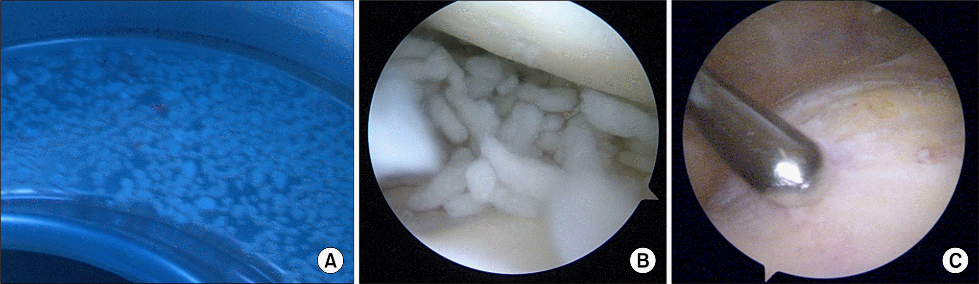 | Figure 2.Gross morphology of rice bodies in the washing synovial fluid and arthroscopic findings. (A) Many white amorphous materials were found in the washing synovial fluid collected through arthroscopic irrigation. Arthroscopic findings were consistent with rice bodies in the suprapatellar space (B) and revealed a normal synovium (C). |
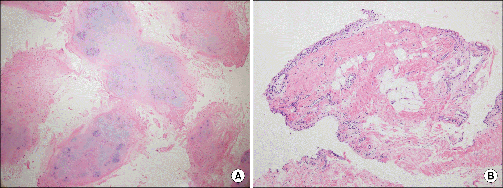 | Figure 3.Pathology of rice body and synovium. (A) Microscopic examination (H&E, x40) revealed multiple nodular fi-brocartilaginous tissues consistent with rice bodies. (B) The synovium of the knee joint (H&E, x100) showed a histologically clean synovial surface. |
Table 1.
Clinical characteristics and prognosis after treatment of idiopathic rice bodies
 XML Download