Abstract
A-43-year-old man visited our clinic due to pain and swelling of his left first metatarsophalangeal (MTP) joint since 6-months ago. He was diagnosed as gouty arthritis at private clinic and took hypouricemic agent, but he had progressive pain and swelling. There was swelling, erythema and tenderness and ulceration at base of the left first MTP joint. His laboratory results showed elevated C-reactive protein and normal serum uric acid level. The plain radiograph of foot showed bone destruction of left first MTP joint. MRI revealed joint space narrowing, soft tissue swelling and subchondral cyst. He underwent excisional biopsy and histology demonstrated chronic granulomatous inflammation with caseation necrosis. Tissue polymerase chain reaction for mycobacterium tuberculosis was positive. He was diagnosed as tuberculous osteomyelitis. He started on quadruple anti-tuberculous therapy and his symptom was improved. Early diagnosis and anti-tuberculosis therapy could lead to improve outcomes.
REFERENCES
1. Korim M, Patel R, Allen P, Mangwani J. Foot and ankle tuberculosis: case series and literature review. Foot (Edinb). 2014; 24:176–9.

2. Samuel S, Boopalan PR, Alexander M, Ismavel R, Varghese VD, Mathai T. Tuberculosis of and around the ankle. J Foot Ankle Surg. 2011; 50:466–72.

4. Gursu S, Yildirim T, Ucpinar H, Sofu H, Camurcu Y, Sahin V, et al. Long-term follow-up results of foot and ankle tuberculosis in Turkey. J Foot Ankle Surg. 2014; 53:557–61.

5. Choi JS, Gwak HC, Kim JH, Chung HJ. Tuberculosis in foot and ankle. J Korean Foot Ankle Soc. 2008; 12:203–9.
6. Choi JS, Gwak HC, Kim JH, Lee CR. Tuberculous osteomyelitis of the tarsal bone in an infant: case report. J Korean Orthop Assoc. 2009; 44:275–8.
7. Nayak B, Dash RR, Mohapatra KC, Panda G. Ankle and foot tuberculosis: a diagnostic dilemma. J Family Med Prim Care. 2014; 3:129–31.

8. Chevannes W, Memarzadeh A, Pasapula C. Isolated tuberculous osteomyelitis of the talonavicular joint without pulmonary involvement-a rare case report. Foot (Edinb). 2015; 25:66–8.

9. Golden MP, Vikram HR. Extrapulmonary tuberculosis: an overview. Am Fam Physician. 2005; 72:1761–8.
10. Parmar H, Shah J, Patkar D, Singrakhia M, Patankar T, Hutchinson C. Tuberculous arthritis of the appendicular skeleton: MR imaging appearances. Eur J Radiol. 2004; 52:300–9.

11. Tsai YJ, Shiau YC. Diagnosis and monitoring treatment response of skeletal tuberculosis of foot by three-phase bone scan: a case report. Ann Nucl Med Sci. 2010; 23:175–80.
12. Hong L, Wu JG, Ding JG, Wang XY, Zheng MH, Fu RQ, et al. Multifocal skeletal tuberculosis: experience in diagnosis and treatment. Med Mal Infect. 2010; 40:6–11.

13. Hsiao CH, Cheng A, Huang YT, Liao CH, Hsueh PR. Clinical and pathological characteristics of mycobacterial tenosynovitis and arthritis. Infection. 2013; 41:457–64.

Figure 1.
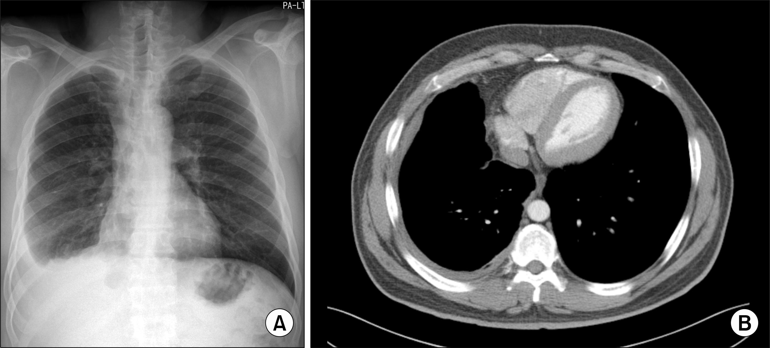
(A) Chest radiograph showed right costophrenic angle blunting. (B) Computed tomography image of chest showed diffuse pleural thickening in right lower lobe.

Figure 2.
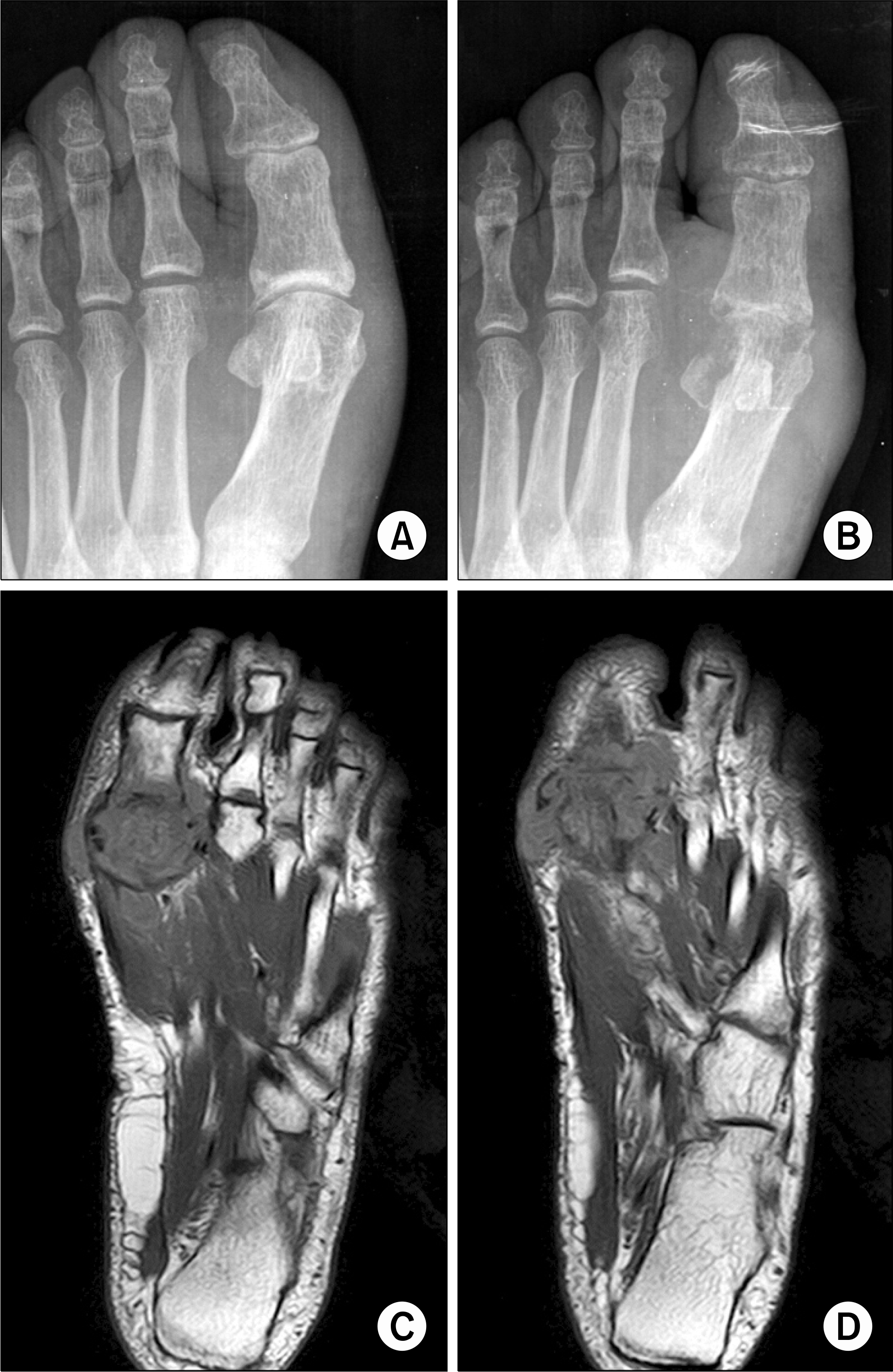
(A) There was no destruction at left first meta-tarsophalangeal (MTP) joint in foot X-ray at 6-months ago. (B) There was bone destruction at left first MTP joint on day of admission. (C, D) Magnetic resonance imaging revealed joint destruction, soft tissue swelling and subchondral erosion in left first MTP joint.

Figure 3.
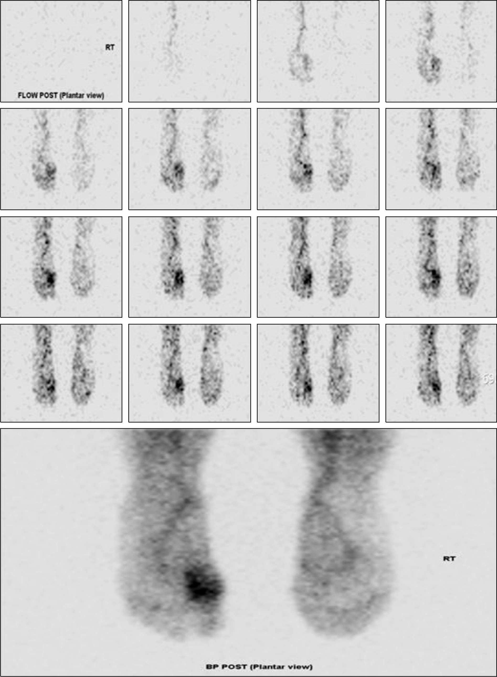
Three phase bone scan image showed increased perfusion over left foot in the dynamic images. In the blood flow and pool phase, there was increased flow in the left first metatarsophalangeal joint area. These findings are compatible with osteomyelitis.

Figure 4.
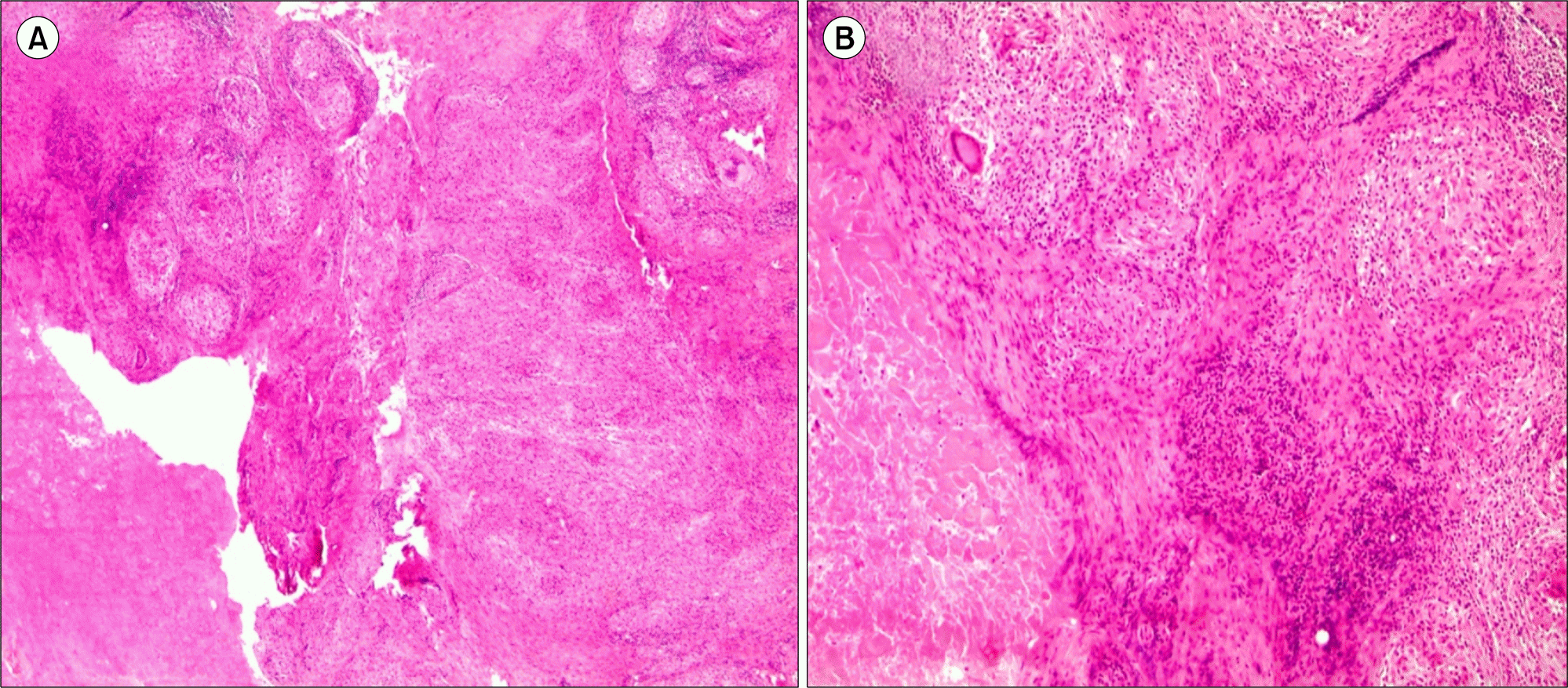
Tissue from excision biopsy of the left first metatarsophalangeal joint showed chronic granulomatous inflammation with caseation necrosis. This histologic finding is compatible with tuberculosis. (H&E: A, ×40; B, ×100).

Table 1.
Summary of foot and ankle tuberc culosis case series


 PDF
PDF ePub
ePub Citation
 Print
Print


 XML Download