

 PDF
PDF ePub
ePub Citation
 Print
Print


Abstract
Polymyositis (PM) is a subset of idiopathic inflammatory myopathies. The muscles involved with PM are typically proximal and distal limb muscles, but paraspinal muscles are rarely affected. The primary PM clinical symptom is gradual proximal muscle weakness but unusually abnormal trunk posture. Bent spine syndrome (BSS), also referred to camptocormia, is defined as an abnormal flexion of the trunk, appearing in standing position. An idiopathic axial myopathy is the most common cause of primary BSS. A few cases of inflammatory myopathy, a secondary BSS, have been reported. We describe a 59-year– old polymyositis patient with normal finding on an magnetic resonance imaging femur scan who presented with BSS only, myopathic findings on electromyography and elevation of muscle enzymes.
REFERENCES
1. Christopher-Stine L, Plotz PH. Adult inflammatory myopathies. Best Pract Res Clin Rheumatol. 2004; 18:331–44.
2. Dalakas MC, Hohlfeld R. Polymyositis and dermatomyositis. Lancet. 2003; 362:971–82.

3. Dalakas MC. Inflammatory muscle diseases. N Engl J Med. 2015; 373:393–4.
4. Lenoir T, Guedj N, Boulu P, Guigui P, Benoist M. Camptocormia: the bent spine syndrome, an update. Eur Spine J. 2010; 19:1229–37.
5. Laroche M, Cintas P. Bent spine syndrome (camptocormia): a retrospective study of 63 patients. Joint Bone Spine. 2010; 77:593–6.
6. Serratrice G, Pouget J, Pellissier JF. Bent spine syndrome. J Neurol Neurosurg Psychiatry. 1996; 60:51–4.
7. Finsterer J, Strobl W. Presentation, etiology, diagnosis, and management of camptocormia. Eur Neurol. 2010; 64:1–8.
8. Kim JM, Song EJ, Seo JS, Nam EJ, Kang YM. Polymyositis – like syndrome caused by hypothyroidism, presenting as camptocormia. Rheumatol Int. 2009; 29:339–42.
9. Dyck PJ, Boes CJ, Mulder D, Millikan C, Windebank AJ, Dyck PJ, et al. History of standard scoring, notation, and summation of neuromuscular signs. A current survey and recommendation. J Peripher Nerv Syst. 2005; 10:158–73.
10. Mahjneh I, Marconi G, Paetau A, Saarinen A, Salmi T, Somer H. Axial myopathy–an unrecognised entity. J Neurol. 2002; 249:730–4.
11. Djaldetti R, Mosberg-Galili R, Sroka H, Merims D, Melamed E. Camptocormia (bent spine) in patients with Parkinson's disease–characterization and possible pathogenesis of an unusual phenomenon. Mov Disord. 1999; 14:443–7.
12. Gdynia HJ, Sperfeld AD, Unrath A, Ludolph AC, Sabolek M, Storch A, et al. Histopathological analysis of skeletal muscle in patients with Parkinson's disease and ‘dropped head’/‘bent spine’ syndrome. Parkinsonism Relat Disord. 2009; 15:633–9.
13. Kuo SH, Vullaganti M, Jimenez-Shahed J, Kwan JY. Camptocormia as a presentation of generalized inflammatory myopathy. Muscle Nerve. 2009; 40:1059–63.
14. Mattar MA, Gordo JM, Halpern AS, Shinjo SK. Camptocormia secondary to polymyositis. Rev Bras Reumatol. 2013; 53:368–70.
15. Delcey V, Hachulla E, Michon-Pasturel U, Queyrel V, Hatron PY, Boutry N, et al. [Camptocormia: a sign of axial myopathy. Report of 7 cases]. Rev Med Interne. 2002; 23:144–54. In French.
Figure 1.
Magnetic resonance imaging scan for thighs at rehospitalization. Axial T2-weighted scan (A) and axial fatsaturated T2-weighted scan (B) revealed bilaterally fatty atrophy of semimembranosus muscle (arrow) without active inflammation.
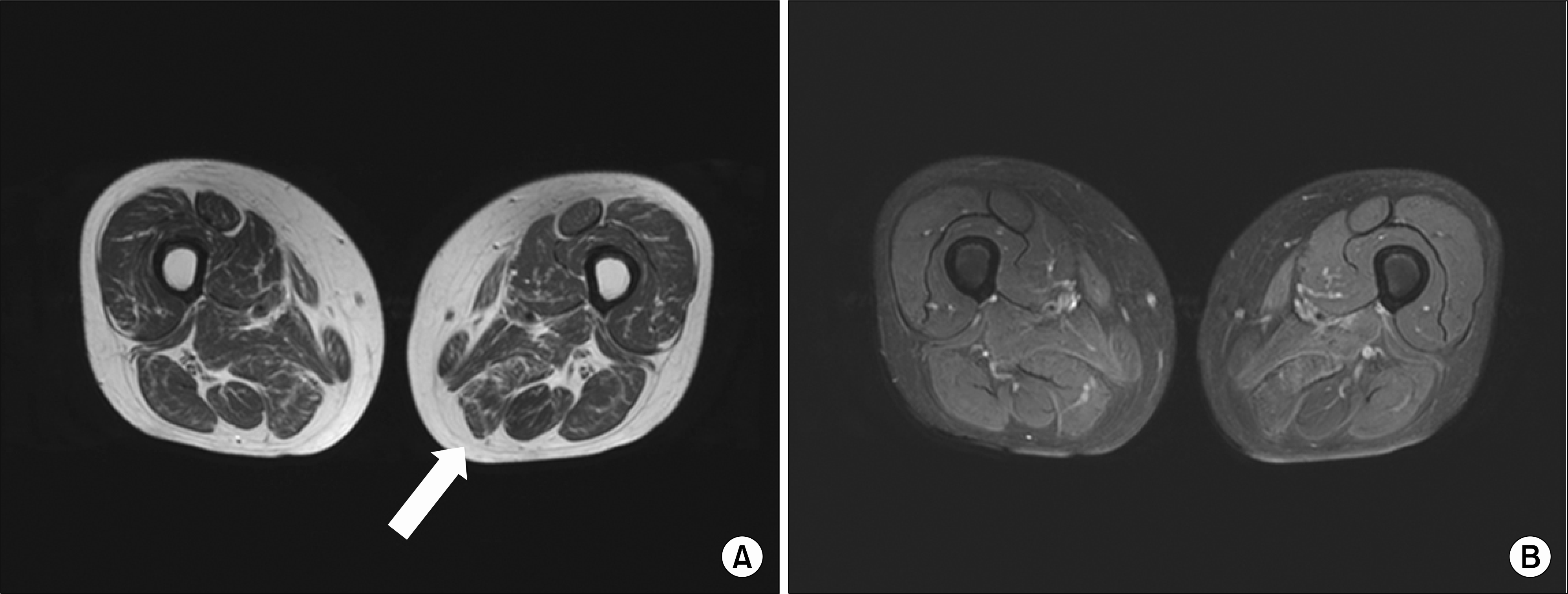
Figure 2.
Axial lumbar spine magnetic resonance imaging. Fat saturated T1-weighted scan (A) and T2-weighted scan (B) showed paraspinous muscle edema with enhancement, especially worse on the right iliocostalis muscle (arrow) and longissimus (arrow head) at the level of L2 and L3.

Figure 3.
Pathologic finding of paraspinal muscle. (A) The right longissimus muscle demonstrates endomysial lymphocytic infiltration (H&E, ×100). (B) CD8 cytotoxic T-cell in the same tissue (immunohistochemical stain, ×200).
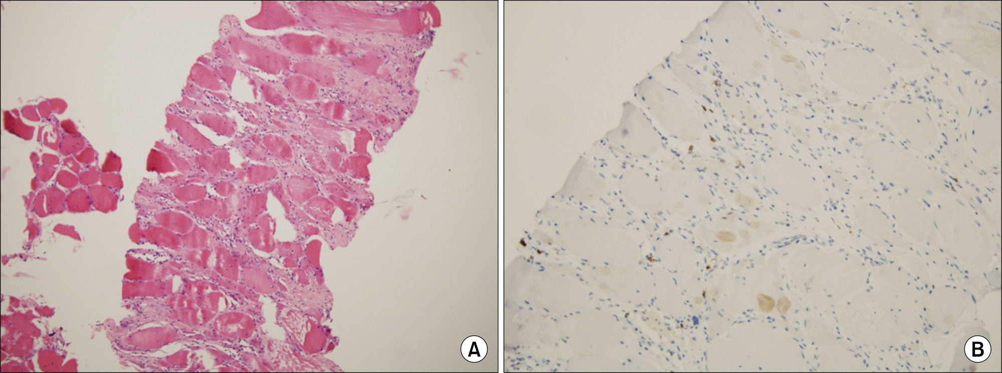
Table 1.
Patient cases of polymyositis with bent spine syndrome
 XML Download