Abstract
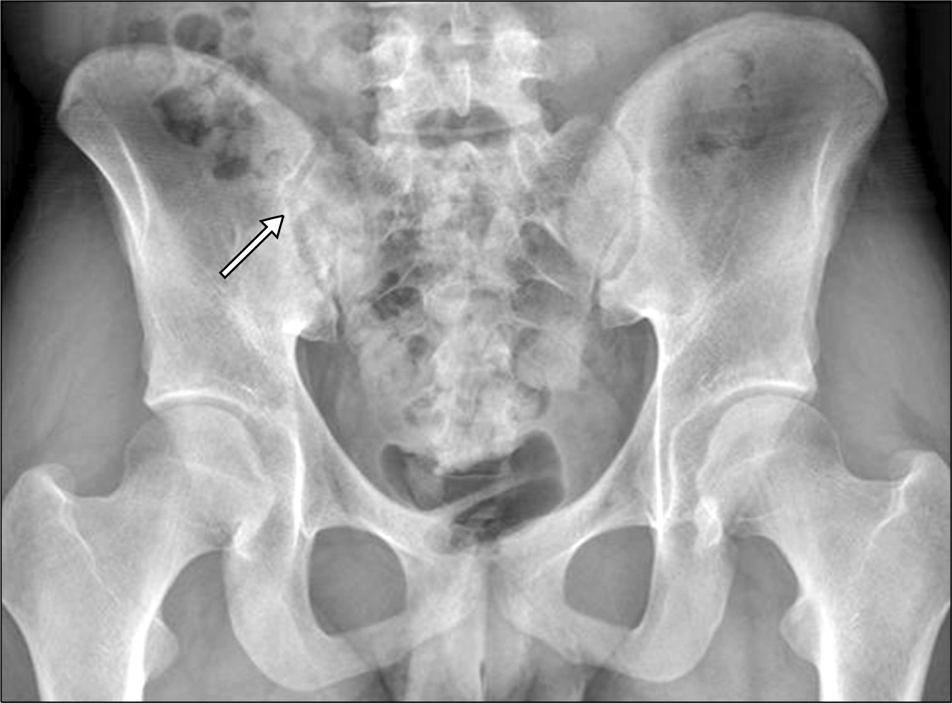
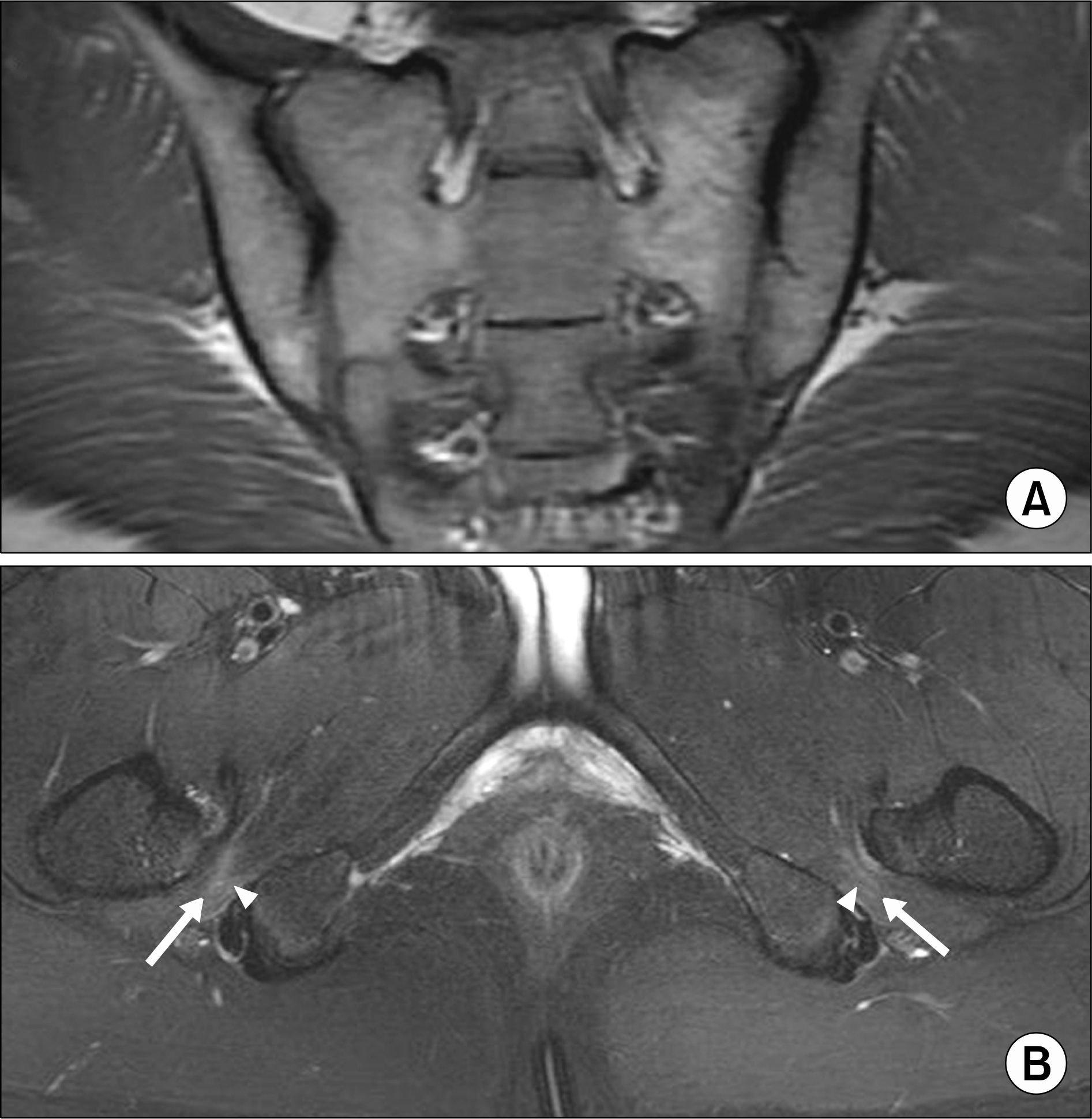
Ischiofemoral impingement (IFI) syndrome is an uncommon cause of gluteal and hip pain. We report on a case of a 20-year-old man who presented with chronic gluteal and hip pain with low back pain without a history of trauma or surgery. He was misdiagnosed with ankylosing spondylitis (AS) at another clinic. The patient was finally diagnosed with IFI syndrome according to pelvic magnetic resonance imaging findings at our hospital. After two weeks of medical and physical treatment, his pain showed gradual improvement. Because IFI syndrome is rarely reported in male patients, it might be misdiagnosed as AS. Therefore, IFI syndrome should be considered as a differential diagnosis of AS, particularly in young male patients with atypical pain characteristics.
REFERENCES
1. Johnson KA. Impingement of the lesser trochanter on the ischial ramus after total hip arthroplasty. Report of three cases. J Bone Joint Surg Am. 1977; 59:268–9.

2. Patti JW, Ouellette H, Bredella MA, Torriani M. Impingement of lesser trochanter on ischium as a potential cause for hip pain. Skeletal Radiol. 2008; 37:939–41.

3. López-Sánchez MC, Armesto Pérez V, Montero Furelos LÁ, Vázquez-Rodríguez TR, Calvo Arrojo G, Díaz Román TM. Ischiofemoral impingement: hip pain of infrequent cause. Reumatol Clin. 2013; 9:186–7.
4. Kim WJ, Shin HY, Koo GH, Park HG, Ha YC, Park YH. Ultrasound-guided prolotherapy with polydeoxyribonu-cleotide sodium in ischiofemoral impingement syndrome. Pain Pract. 2014; 14:649–55.

5. Singer AD, Subhawong TK, Jose J, Tresley J, Clifford PD. Ischiofemoral impingement syndrome: a meta-analysis. Skeletal Radiol. 2015; 44:831–7.

6. Safran M, Ryu J. Ischiofemoral impingement of the hip: a novel approach to treatment. Knee Surg Sports Traumatol Arthrosc. 2014; 22:781–5.

7. Papavasiliou A, Siatras T, Bintoudi A, Milosis D, Lallas V, Sykaras E, et al. The gymnasts' hip and groin: a magnetic resonance imaging study in asymptomatic elite athletes. Skeletal Radiol. 2014; 43:1071–7.

8. Torriani M, Souto SC, Thomas BJ, Ouellette H, Bredella MA. Ischiofemoral impingement syndrome: an entity with hip pain and abnormalities of the quadratus femoris muscle. AJR Am J Roentgenol. 2009; 193:186–90.

9. Yazici H, Turunç M, Ozdoğan H, Yurdakul S, Akinci A, Barnes CG. Observer variation in grading sacroiliac radiographs might be a cause of ‘sacroiliitis’ reported in certain disease states. Ann Rheum Dis. 1987; 46:139–45.

10. Cohen AS, McNeill JM, Calkins E, Sharp JT, Schubart A. The “normal” sacroiliac joint. Analysis of 88 sacroiliac roentgenograms. Am J Roentgenol Radium Ther Nucl Med. 1967; 100:559–63.
11. Oostveen J, Prevo R, den Boer J, van de Laar M. Early detection of sacroiliitis on magnetic resonance imaging and subsequent development of sacroiliitis on plain radiography. A prospective, longitudinal study. J Rheumatol. 1999; 26:1953–8.


 PDF
PDF ePub
ePub Citation
 Print
Print


 XML Download