Journal List > J Rheum Dis > v.23(2) > 1064254

Park and Chae: Spontaneous Achilles Tendon Rupture in a Patient with Ankylosing Spondylitis

REFERENCES

1. Braun J, Sieper J. Ankylosing spondylitis. Lancet. 2007; 369:1379–90.
crossref
2. Kim HW, Lee SH. Pathogenesis of ankylosing spondylitis. J Rheum Dis. 2015; 22:61–8.
crossref
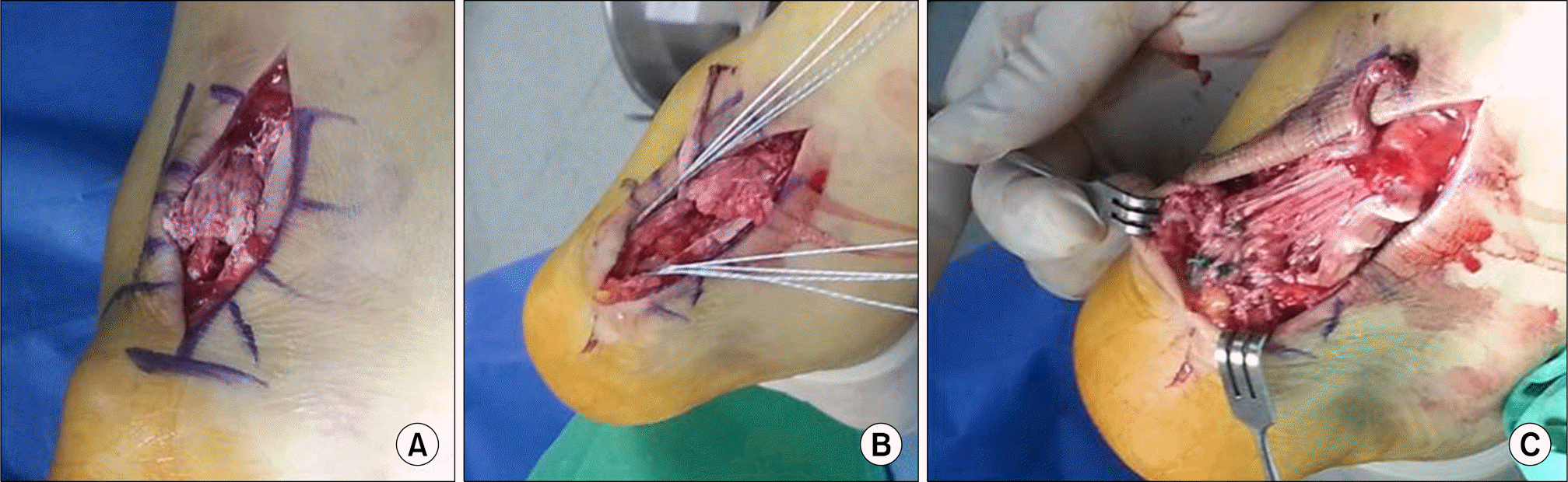
3. Jiang N, Wang B, Chen A, Dong F, Yu B. Operative versus nonoperative treatment for acute Achilles tendon rupture: a meta-analysis based on current evidence. Int Orthop. 2012; 36:765–73.
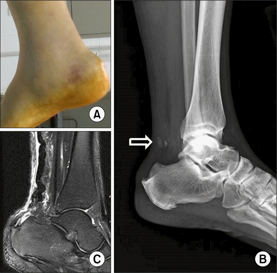
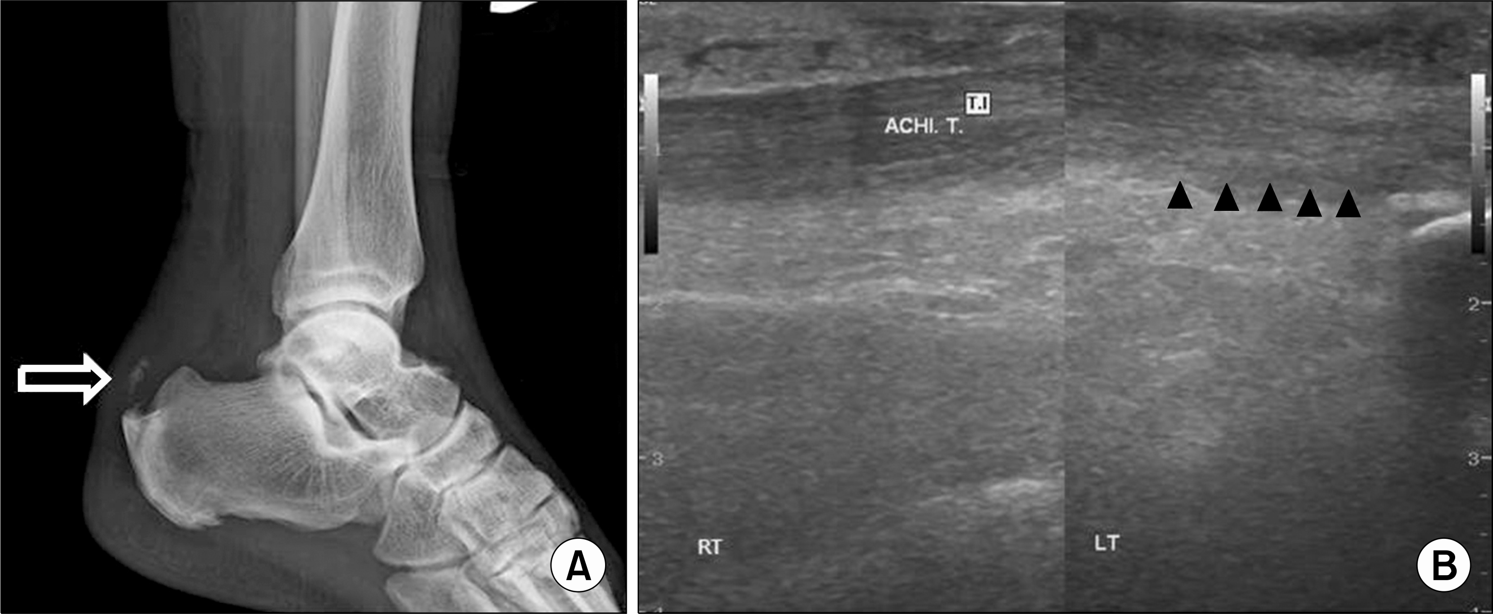
crossref

Figure 1.
Simple anteroposterior (A) and lateral (B) L-spine radiography show a bamboo spine.
jrd-23-136f1.tif
Figure 2.
(A) The clinical photo shows swelling, ecchymosis and dimpling in the posterior aspect of the ankle. (B) Simple lateral ankle image shows loss of Kager's triangle and bony fragments (arrow). (C) Sagittal T2 magnetic resonance image shows rupture of the Achilles tendon at calcaneal insertion site and enthesopathic spur.
jrd-23-136f2.tif
Figure 3.
(A) Intraoperative finding shows the rupture of the Achilles tendon at calcaneal insertion site and combined with bony fragments. (B) Achilles tendon rupture was treated with tendon to bone repair using suture anchors. (C) Postoperative finding shows complete repair tendon to bone repair.
jrd-23-136f3.tif
Figure 4.
Six months follow-up simple lateral ankle image shows intact morphology of Kager's triangle (arrow) (A), ultrasonography shows normal distal Achilles tendon with fibrillar pattern at calcaneal insertion (arrowheads) (B). ACHI. T: Achilles tendon, LT: left, RT: right.
jrd-23-136f4.tif
TOOLS
Similar articles