

 PDF
PDF ePub
ePub Citation
 Print
Print


Abstract
Calcium pyrophosphate dihydrate crystal deposition disease is associated with an acute mono- or pauciarthritis, termed “pseudogout” in elderly patients, involving a large joint (including the knees, ankles) or a chronic arthropathy manifesting as mild joint pain and stiffness. Pseudogout is a crystal-deposition disease of peripheral joints, usually encountered in elderly patients. However, acute presentation of pseudogout around the odontoid process comprises a “crowned-dens” appearance, and requires contemplation of differential diagnoses. We recently experienced a case of pseudogout in the cervical spine presenting with fever and acute neck pain that was successfully treated with a colchicine and low-dose oral steroid. We reported this case with a review of the relevant literature.
Go to : 
REFERENCES
1. Wise CM. Crystal-associated arthritis in the elderly. Rheum Dis Clin North Am. 2007; 33:33–55.

2. Abhishek A, Doherty M. Epidemiology of calcium pyrophosphate crystal arthritis and basic calcium phosphate crystal arthropathy. Rheum Dis Clin North Am. 2014; 40:177–91.
3. Bouvet JP, le Parc JM, Michalski B, Benlahrache C, Auquier L. Acute neck pain due to calcifications surrounding the odontoid process: the crowned dens syndrome. Arthritis Rheum. 1985; 28:1417–20.
4. Jeon CH, Choe WH, Ahn JK, Koh JH, Cha HS, Ahn JM, et al. Calcium pyrophosphate dihydrate (CPPD) crystal deposition disease mimicking meningitis: A case report and review of the literature. J Korean Rheum Assoc. 2001; 8:134–9.
5. Ishikawa K, Furuya T, Noda K, Okuma Y. Crowned dens syndrome mimicking meningitis. Intern Med. 2010; 49:2023.
6. Mahmud T, Basu D, Dyson PH. Crystal arthropathy of the lumbar spine: a series of six cases and a review of the literature. J Bone Joint Surg Br. 2005; 87:513–7.
7. Finckh A, Van Linthoudt D, Duvoisin B, Bovay P, Gerster JC. The cervical spine in calcium pyrophosphate dihydrate deposition disease. A prevalent case-control study. J Rheumatol. 2004; 31:545–9.
8. Viana SL, Fernandes JL, De Araújo Coimbra PP, De Mendonça JL, Freitas FM, De Carvalho Barbosa Viana MA. The “crowned dens” revisited: imaging findings in calcium crystal deposition diseases around the odontoid. J Neuroimaging. 2010; 20:311–23.
9. Koyfman A, Yaffe D. Crowned dens syndrome. A case report. Neuroradiol J. 2014; 27:495–7.
10. Zhang W, Doherty M, Pascual E, Barskova V, Guerne PA, Jansen TL, et al. EULAR recommendations for calcium pyrophosphate deposition. Part II: management. Ann Rheum Dis. 2011; 70:571–5.
11. Nuki G. Colchicine: its mechanism of action and efficacy in crystal-induced inflammation. Curr Rheumatol Rep. 2008; 10:218–27.
12. Sethi KS, Garg A, Sharma MC, Ahmad FU, Sharma BS. Cervicomedullary compression secondary to massive calcium pyrophosphate crystal deposition in the atlantoaxial joint with intradural extension and vertebral artery encasement. Surg Neurol. 2007; 67:200–3.
Go to :
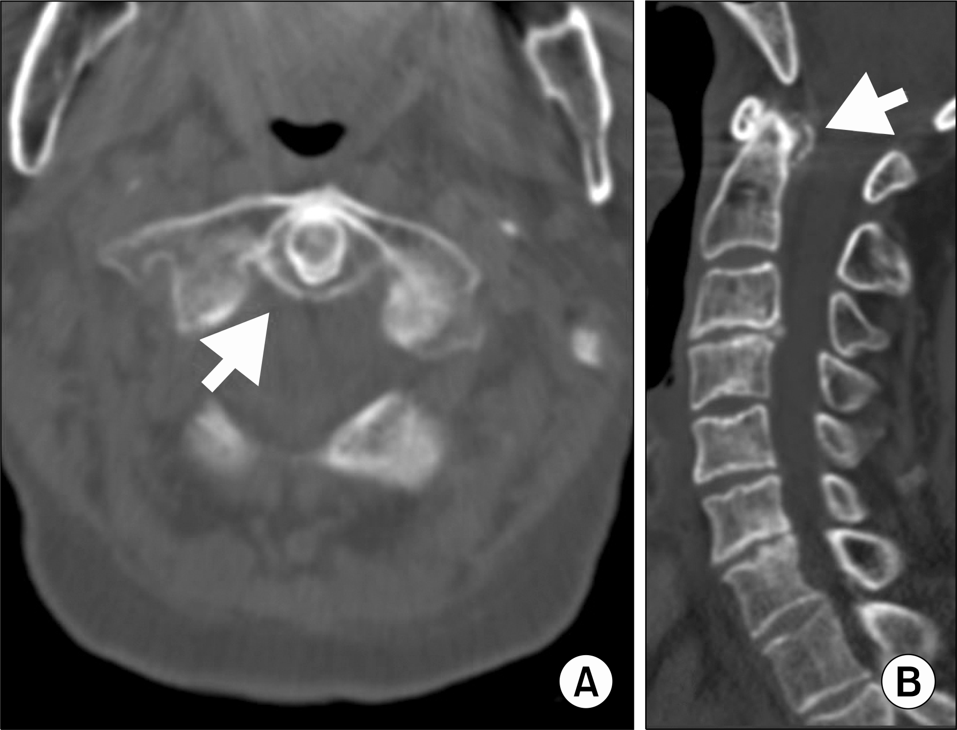 | Figure 1.Axial (A) and sagittal (B) image of the cervical computed tomography scan at the C1/C2 level shows curvilinear calcifications of the transverse ligament (arrows). |
 XML Download