Abstract
Psoriatic onycho-pachydermo-periostitis (POPP) causes severe nail dystrophy, painful soft tissue swelling, and marked periosteal reaction of the involved distal phalanx. There are few reports of POPP involving the great toe. We report on 2 cases of POPP involving the fingertips. A 60-year-old woman presented with fusiform swelling of her right 4th fingertip with severe tenderness, and her fingernails and toenails had varying degrees of onycholysis. She had mixed multiple erosions and meta-epi-physeal periostitis at the distal phalanx of the right 4th finger but was treated successfully with methotrexate and cyclosporine. A 39-year-old woman presented with painful swelling of the left 2nd and 5th fingertip, psoriatic lesions on the knees and soles of the feet, and onycholysis without reactive periostitis of the left 2nd and 5th fingers. She was treated successfully with cyclosporine. Despite its rarity, POPP should be considered when diagnosing arthritic or infectious conditions affecting the distal interphalangeal joint.
REFERENCES
1. Fournié B, Viraben R, Durroux R, Lassoued S, Gay R, Fournié A. Psoriatic onycho-pachydermo-periostitis of the big toe. Anatomo-clinical study and physiopathogenic approach apropos of 4 cases. Rev Rhum Mal Osteoartic. 1989; 56:579–82.
2. Marguery MC, Baran R, Pages M, Bazex J. Psoriatic acropachydermy. Ann Dermatol Venereol. 1991; 118:373–6.
3. De Pontville M, Dompmartin A, De Raucourt S, Macro M, Rémond B, Leroy D. Psoriatic onycho-pachydermo-periostitis. Ann Dermatol Venereol. 1993; 120:229–32.
4. Grosshans E, Bosser V. A case for diagnosis: ungueal psoriasis. Ann Dermatol Venereol. 1993; 120:319–20.
5. Boisseau-Garsaud AM, Beylot-Barry M, Doutre MS, Beylot C, Baran R. Psoriatic onycho-pachydermo-periostitis. A variant of psoriatic distal interphalangeal arthritis? Arch Dermatol. 1996; 132:176–80.

6. Bauzá A, Redondo P, Aquerreta D. Psoriatic onycho-pachy-dermo periostitis: treatment with methotrexate. Br J Dermatol. 2000; 143:901–2.

7. Anders HJ, Sanden S, Krüger K. Psoriatic onychopachy-dermoperiostitis. Z Rheumatol. 2002; 61:601–4.
8. Fietta P, Manganelli P. Pachydermoperiostosis and psoriatic onychopathy: an unusual association. J Eur Acad Dermatol Venereol. 2003; 17:73–6.

9. Bongartz T, Härle P, Friedrich S, Karrer S, Vogt T, Seitz A, et al. Successful treatment of psoriatic onycho-pachydermo periostitis (POPP) with adalimumab. Arthritis Rheum. 2005; 52:280–2.

10. Kapusta MA, Dumont C. Differential response of psoriatic onycho-pachydermo-periostitis to 2 antitumor necrosis factor-alpha agents. J Rheumatol. 2008; 35:2077–80.
11. Watanabe M, Ujiie H, Iitani MM, Abe R, Shimizu H. Psoriatic onycho-pachydermo-periostitis progressing to generalized pustular psoriasis. Clin Exp Dermatol. 2012; 37:683–5.

12. Bethapudi S, Halstead J, Ash Z, McGonagle D, Grainger AJ. Test yourself: answer psoriatic onycho-pachydermo periostitis (POPP). Skeletal Radiol. 2014; 43:409–11.

14. Koó T, Nagy Z, Seszták M, Ujfalussy I, Merétey K, Böhm U, et al. Subsets in psoriatic arthritis formed by cluster analysis. Clin Rheumatol. 2001; 20:36–43.

15. Ingram GJ, Scher RK. Reiter' s syndrome with nail involvement: is it psoriasis? Cutis. 1985; 36:37–40.
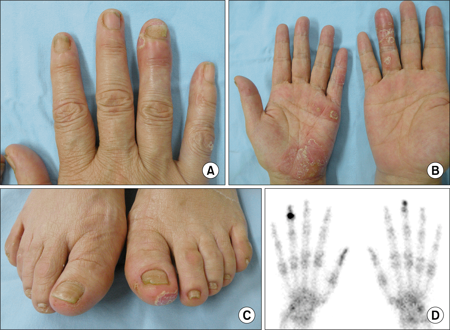
Figure 1.
The first patient's skin lesion and three-phase bone scintigraph. (A∼ C) The right 4th fingertip shows fusiform swelling with erythema, and the nails of the fingers and toes demonstrate dystrophic onycholysis. (B, C) Pustular psoriatic lesions are noted in the left palm and right great toe. (D) Three-phase bone scintigraphy shows a focal radio-nuclide accumulation in the right 4th and left 3rd distal phalanx fingers on a delayed static scan.

Figure 2.
A serial change of the hand is shown on anteroposterior radiographs. (A) The radiograph taken 6 months prior to presentation demonstrates meta-epi-physeal periostitis and multiple erosions of the distal phalanx of the left 4th finger. (B) After 4 months, a reactive bone formation is observed at the shaft of the distal phalanx (arrow). (C) The radiograph at presentation reveals progression of erosion and marked soft tissue swelling (arrow).

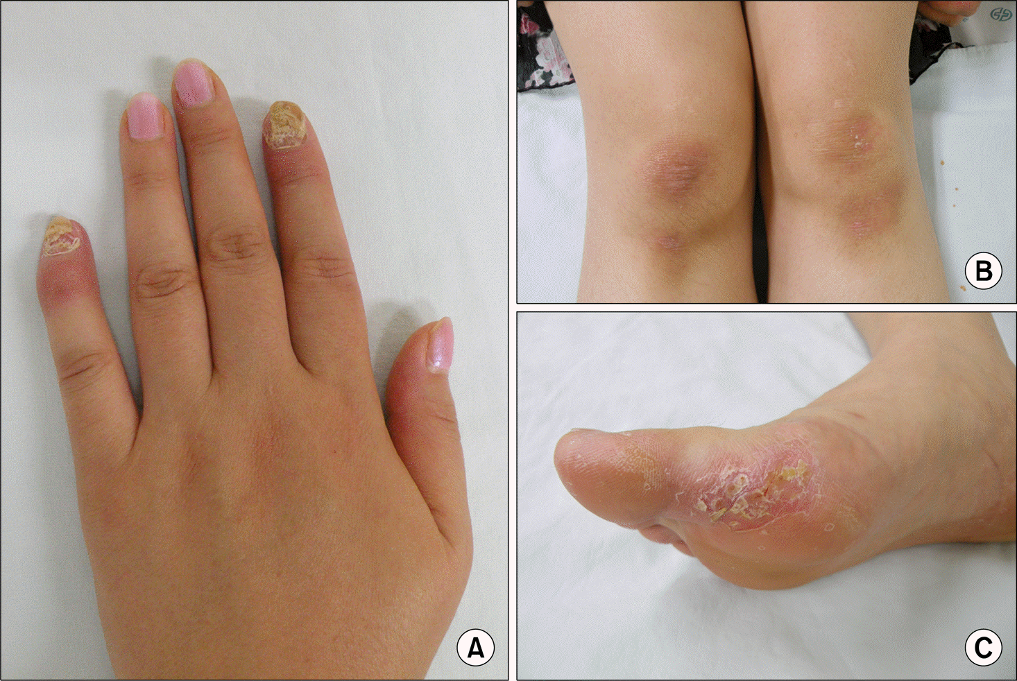
Figure 3.
The second patient's hand and skin lesions before treatment. (A) The left 2nd and 5th fingers exhibit drumstick-like swelling and inflammation, and the fingernails show severe onycholysis. (B, C) Erythematous, hyperkeratotic plaque is seen on both knee and the sole of the right foot.

Table 1.
Summary of clinical characteristics of the 21 psoriatic onycho-pachydermo-periostitis patients
| Study | Age (yr) | Sex | Disease duration | Skin psoriasis | Onycholysis | Distal phalanx swelling | HLA-B27 | Treatment |
|---|---|---|---|---|---|---|---|---|
| Fournié et al. [1] (1989)* | 57 | NA | 2 yr | None | Both 1st toes | None | Positive | NA |
| 45 | NA | 2 yr | None | Both 1st toes, left 3rd finger | None | NA | NA | |
| 46 | NA | 20 yr | None | Both 1st toes | None | NA | NA | |
| 39 | NA | 3 yr | None | Right 1st toe | None | NA | NA | |
| Marguery et al. [2](1991)* | 55 | M | 2∼3 yr | NA | All fingers except left 5th finger | Right 3rd, 4th and left 2∼4th fingers | NA | NA |
| De Pontville et al. [3] (1993)* | 40 | NA | 1 yr | NA | All fingers and toes | Right 1st finger, right 1st, 3rd, 4th toe, left 1st, 4th toe | Positive | NA |
| 62 | NA | 1 yr | NA | Right 1st toe | None | Positive | NA | |
| Grosshans and Bosser [4] (1993)* | 35 | NA | Several years | NA | Left 1st toe | None | NA | NA |
| 37 | NA | 2 yr | NA | Left 3rd finger, both 1st toes | Left 3rd finger | Positive | NA | |
| 49 | NA | 5 yr | NA | Right 1st finger | None | NA | NA | |
| Boisseau-Garsaud et al. [5] (1996) | 37 | M | 2 yr | None | Left 1st∼5th fingers, both 1st toes | Left 2nd and 3rd fingers, both 1st toes | Positive | MTX 20 mg/wk (response) |
| 49 | M | 5 yr | Both elbow, scalp | Right 1st finger | Right 1st finger | NA | NA | |
| Bauzá et al. [6] (2000) | 50 | F | 2 yr | None | Left 3rd∼5th fingers, right 3rd finger | Both 3rd fingers | NA | Cyclosporin A 200 mg→ MTX 15 mg/wk |
| Anders et al. [7] (2002) | 49 | M | NA | None | All fingers and toes | Right 3rd, 4th and left 2nd and 3rd fingers, left 1st toe | NA | SSZ→ radiotherapy |
| Fietta and Manganelli [8] (2003) | 33 | M | 6 yr | Both elbows, leg | All fingers | None | Negative | Topical steroid |
| Bongartz et al. [9] (2005) | 42 | M | 1.5 yr | Both feet | All toes | All toes | Positive | MTX 15 mg/wk→ adalimumab |
| Kapusta and Dumont [10] (2008) | 53 | M | 0.5 yr | Right ear, both feet | All fingers and toes | Left 1st, 2nd, and 4th toes; both 2nd, 3rd, and 5th fingers | Negative | MTX→ etanercept→ infliximab |
| Watanabe et al. [11] (2012) | 59 | M | 3 mo | Whole body | All fingers and toes | All fingers and toes | Negative | Etretinate→ MTX 8 mg/wk |
| Bethapudi et al. [12] (2014) | 55 | F | 1 yr | Palm, foot | All toes | Right 1st, 4th toe, left 1st toe | NA | NA |
| This study (2014) | 60 | F | 4 yr | Both hands, foot | All fingers and toes | Left 4th finger | Negative | MTX, cyclosporin A |
| 39 | F | 1 yr | Both knees, right foot | Left 2nd and 5th fingers | Left 2nd and 4th fingers | Negative | Cyclosporin A |


 PDF
PDF ePub
ePub Citation
 Print
Print


 XML Download