Abstract
Cytomegalovirus (CMV), a member of the human herpesvirus group, causes severe disease in immunocompromised patients. In particular, CMV pneumonia can be a life-threatening disease to patients taking immunosuppressive drugs. The radiographic manifestations of CMV are variable and may consist of reticular or reticulonodular patterns, ground-glass opacities, air-space consolidations, or mixed patterns. A cavitary lesion in pneumonia associated with CMV infection is extremely rare. Herein we report on a case of CMV pneumonia which presented with a cavitary lesion and was treated successfully in a systemic lupus erythematosus patient who was taking immunosuppressive drugs.
Go to : 
REFERENCES
1. Juárez M, Misischia R, Alarcón GS. Infections in systemic connective tissue diseases: systemic lupus erythematosus, scleroderma, and polymyositis/dermatomyositis. Rheum Dis Clin North Am. 2003; 29:163–84.

2. Leland DS, Emanuel D. Laboratory diagnosis of viral infections of the lung. Semin Respir Infect. 1995; 10:189–98.
3. Kang EY, Patz EF Jr, Müller NL. Cytomegalovirus pneumonia in transplant patients: CT findings. J Comput Assist Tomogr. 1996; 20:295–9.

5. Najjar M, Siddiqui AK, Rossoff L, Cohen RI. Cavitary lung masses in SLE patients: an unusual manifestation of CMV infection. Eur Respir J. 2004; 24:182–4.

6. Karakelides H, Aubry MC, Ryu JH. Cytomegalovirus pneumonia mimicking lung cancer in an immunocompetent host. Mayo Clin Proc. 2003; 78:488–90.

7. Katagiri A, Ando T, Kon T, Yamada M, Iida N, Takasaki Y. Cavitary lung lesion in a patient with systemic lupus erythematosus: an unusual manifestation of cytomegalovirus pneumonitis. Mod Rheumatol. 2008; 18:285–9.

8. Azuma N, Hashimoto N, Yasumitsu A, Fukuoka K, Yokoya-ma K, Sawada H, et al. CMV infection presenting as a cavitary lung lesion in a patient with systemic lupus erythematosus receiving immunosuppressive therapy. Intern Med. 2009; 48:2145–9.

9. Lee DH, Kim JW, Shin DH, Oh MD, Song YW, Choi KW, et al. A case of cytomegalovirus pneumonitis in a patient with systemic lupus erythematosus. Korean J Med. 1999; 56:103–7.
10. Han SH, Sohn YJ, Park MA, Lee S, Ryu SH, Lim TH, et al. A case of cytomegalovirus pneumonia and retinitis in a patients with systemic lupus erythematosus. J Korean Rheum Assoc. 2003; 10:456–61.
Go to : 
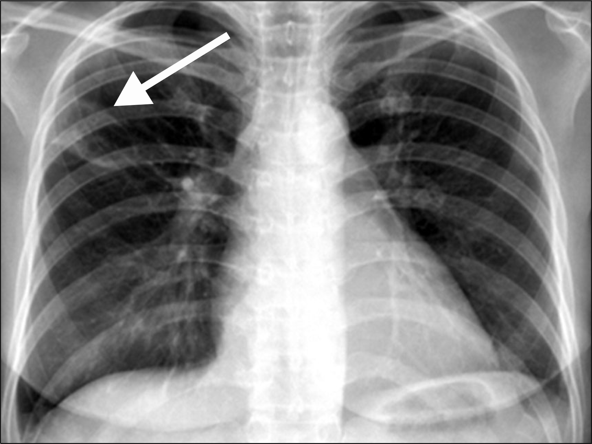
 | Figure 1.Chest plain radiography shows a large cavitary mass like lesion in the right upper lung field (arrow). |
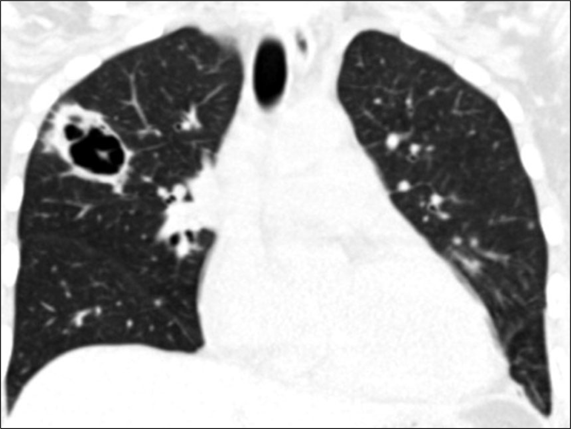
 | Figure 2.Chest computed tomography with coronal reformatted image shows a large cavitary mass like lesion in the right upper lobe. |
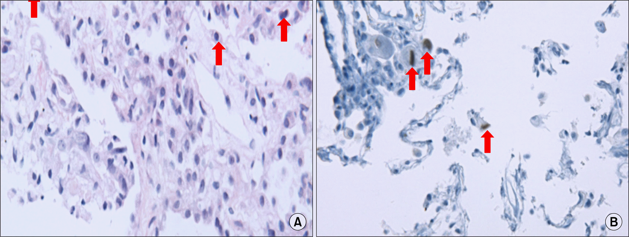
 | Figure 3.(A) The irregularly dilated alveoli showed mononuclear inflammatory cell infiltration in the interstitium and multiple intranuclear cytomegalovirus inclusions in the alveolar pneumonocytes (arrows) (H&E, ×400). (B) Immunohistochemistry (IHC) with an-ti-cytomegalovirus (CMV) confirmed the CMV infected pneumonocytes with intranuclear inclusions (arrows) (IHC, ×400). |


 PDF
PDF ePub
ePub Citation
 Print
Print


 XML Download