Abstract
Behçet's disease (BD) is characterized by recurrent oro-genital ulcers, skin lesions, and intraocular inflammation, but can also affect various internal organs. Vascular BD usually presents with luminal stenosis, thrombosis, or aneurysm formation in aorta and peripheral arteries. However, intracranial artery involvement has been uncommonly reported in patients with BD and BD cases with lenticulostriate artery aneurysm have been rarely described in the English-language literature. We hereby reported the first case of a Korean BD patient presenting with a ruptured lenticulostriate artery aneurysm, who received medical treatment, and reviewed the literature on reported cases of BD with intracranial aneurysms.
REFERENCES
1. Hirohata S, Kikuchi H. Behçet's disease. Arthritis Res Ther. 2003; 5:139–46.
2. Calamia KT, Schirmer M, Melikoglu M. Major vessel involvement in Behçet's disease: an update. Curr Opin Rheumatol. 2011; 23:24–31.

3. Bang D, Lee JH, Lee ES, Lee S, Choi JS, Kim YK, et al. Epidemiologic and clinical survey of Behçet's disease in Korea: the first multicenter study. J Korean Med Sci. 2001; 16:615–8.

4. Al-Araji A, Kidd DP. Neuro-Behçet's disease: epidemiology, clinical characteristics, and management. Lancet Neurol. 2009; 8:192–204.

5. Jeon TY, Jeon P, Kim KH. Prevalence of unruptured intracranial aneurysm on MR angiography. Korean J Radiol. 2011; 12:547–53.

6. Rinne J, Hernesniemi J, Niskanen M, Vapalahti M. Analysis of 561 patients with 690 middle cerebral artery aneurysms: anatomic and clinical features as correlated to management outcome. Neurosurgery. 1996; 38:2–11.

7. Vargas J, Walsh K, Turner R, Chaudry I, Turk A, Spiotta A. Lenticulostriate aneurysms: a case series and review of the literature. J Neurointerv Surg. 2015; 7:194–201.

8. Park EM, Seong JJ, Lee JJ, Kang DW, Roh JK. 2 cases of vas-culo-Behçet's disease involving intracranial artery. J Korean Neurol Assoc. 1999; 17:183–6.
9. Kim TY, Lee JW, Huh SK, Lee KC. Cerebral aneursyms associated with the Behcet's disease. Korean J Cerebrovasc Surg. 2006; 8:135–7.
10. Nakasu S, Kaneko M, Matsuda M. Cerebral aneurysms associated with Behçet's disease: a case report. J Neurol Neurosurg Psychiatry. 2001; 70:682–4.
11. Davatchi F, Shahram F, Chams-Davatchi C, Shams H, Nadji A, Akhlaghi M, et al. Behçet's disease: from East to West. Clin Rheumatol. 2010; 29:823–33.
12. International Study Group for Behçet's Disease. Criteria for diagnosis of Behçet's disease. Lancet. 1990; 335:1078–80.
Figure 1.
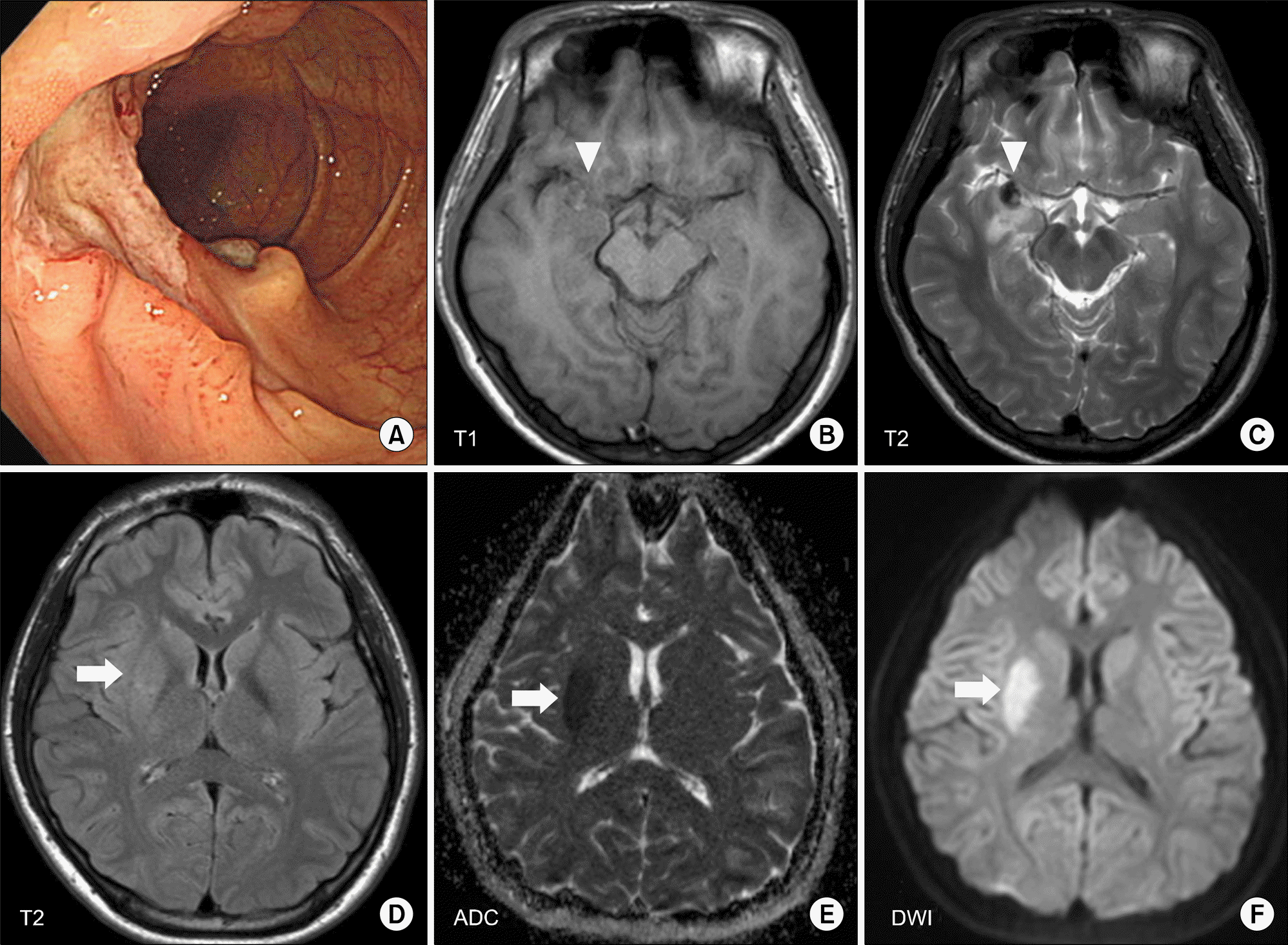
Colonoscopic findings and brain magnetic resonance imaging (MRI) scan images. Colonoscopy showed a large and deep ulcer with a discrete margin in the ileocecal area (A). Acute intracerebral hemorrhage (arrow heads) was visualized in the right temporal pole on axial T1-weighted (B) and T2-weighted (C) scans. An acute infarct in the right basal ganglia (arrows) was clearly identified on axial T2-weighted (D), apparent diffusion coefficient (ADC) (E), and diffusion weighted imaging (DWI) (F) MRI scans.

Figure 2.
Cerebral angiography. Time-of-flight (TOF) magnetic resonance angiography (MRA) using sensitivity encoding (SEN-SE) showed intracerebral hemorrhage adjacent to the M1 segment of the middle cerebral artery (arrow) (A), but MRA did not reveal any abnormality in the M1 segment (B). The initial trans-femoral cerebral angiography (TFCA) prior to glucocorticoid therapy showed a fusiform aneurysm of the right lenticulostriate artery (LSA) and irregu-larity of the vascular wall (C); the inset shows the magnified image of LSA aneurysm (asterisk). After a month of high-dose glucocorticoid treatment, the LSA aneurysm was disappeared on follow-up TFCA (D). ICA: internal carotid artery.

Table 1.
Comparison ofclinical features in Korean Behçet's disease patients with intracranial aneurysm


 PDF
PDF ePub
ePub Citation
 Print
Print


 XML Download