

 PDF
PDF ePub
ePub Citation
 Print
Print


Abstract
Human immunodeficiency virus (HIV) infection is a global pandemic affecting more than 2.9 million people. Aside from opportunistic infections and malignancies, it involves multiple organs, resulting in many complications, and frequently shows various rheumatic manifestations. With improving survival of patients due to the development of highly active anti-retroviral therapy, the number of HIV-infected patients with rheumatic complications is certain to increase. However, reports on HIV induced rheumatic manifestations in Korean patients are limited. On the other hand, spondyloarthropathy is the most common form of inflammatory arthropathy in HIV associated rheumatic manifestations and is frequently accompanied by peripheral arthritis and enthesitis, while axial skeletal involvement is a rare presentation. Herein we report on a 46-year-old man with HIV infection presenting with an axial spondyloarthropathy who was treated successfully with nonsteroidal anti-inflammatory drug, sulfasalazine, and low dose steroid.
Go to : 
REFERENCES
1. Maganti RM, Reveille JD, Williams FM. Therapy insight: the changing spectrum of rheumatic disease in HIV infection. Nat Clin Pract Rheumatol. 2008; 4:428–38.

2. Cuellar ML, Espinoza LR. Rheumatic manifestations of HIV-AIDS. Baillieres Best Pract Res Clin Rheumatol. 2000; 14:579–93.
3. Murray CJ, Ortblad KF, Guinovart C, Lim SS, Wolock TM, Roberts DA, et al. Global, regional, and national incidence and mortality for HIV, tuberculosis, and malaria during 1990-2013: a systematic analysis for the Global Burden of Disease Study 2013. Lancet. 2014; 384:1005–70.
4. Zhang X, Li H, Li T, Zhang F, Han Y. Distinctive rheumatic manifestations in 98 patients with human immunodeficiency virus infection in China. J Rheumatol. 2007; 34:1760–4.
5. Louthrenoo W. Rheumatic manifestations of human immunodeficiency virus infection. Curr Opin Rheumatol. 2008; 20:92–9.
6. Yang JJ, Tsai MS, Sun HY, Hsieh SM, Chen MY, Sheng WH, et al. Autoimmune diseases-related arthritis in HIV-infected patients in the era of highly active antiretroviral therapy. J Microbiol Immunol Infect. 2015; 48:130–6.
7. Iordache L, Launay O, Bouchaud O, Jeantils V, Goujard C, Boue F, et al. Associated authors. Autoimmune diseases in HIV-infected patients: 52 cases and literature review. Autoimmun Rev. 2014; 13:850–7.
8. Ntsiba H, Lamini N. Is inflammatory joint disease in HIV-infected patients a form of spondyloarthropathy? Joint Bone Spine. 2004; 71:300–2.
9. Reveille JD. The changing spectrum of rheumatic disease in human immunodeficiency virus infection. Semin Arthritis Rheum. 2000; 30:147–66.
10. Walker UA, Tyndall A, Daikeler T. Rheumatic conditions in human immunodeficiency virus infection. Rheumatology (Oxford). 2008; 47:952–9.
11. Nguyen BY, Reveille JD. Rheumatic manifestations associated with HIV in the highly active antiretroviral therapy era. Curr Opin Rheumatol. 2009; 21:404–10.
12. Lawson E, Walker-Bone K. The changing spectrum of rheumatic disease in HIV infection. Br Med Bull. 2012; 103:203–21.
13. Clark MR, Solinger AM, Hochberg MC. Human immunodeficiency virus infection is not associated with Reiter's syndrome. Data from three large cohort studies. Rheum Dis Clin North Am. 1992; 18:267–76.
14. Chiowchanwisawakit P, Koolvisoot A, Ratanasuwan W, Suwanagool S. Prevalence of rheumatic disease in HIV infected Thai patients. J Med Assoc Thai. 2005; 88:1775–81.
15. Maurer TA, Zackheim HS, Tuffanelli L, Berger TG. The use of methotrexate for treatment of psoriasis in patients with HIV infection. J Am Acad Dermatol. 1994; 31:372–5.
Go to :
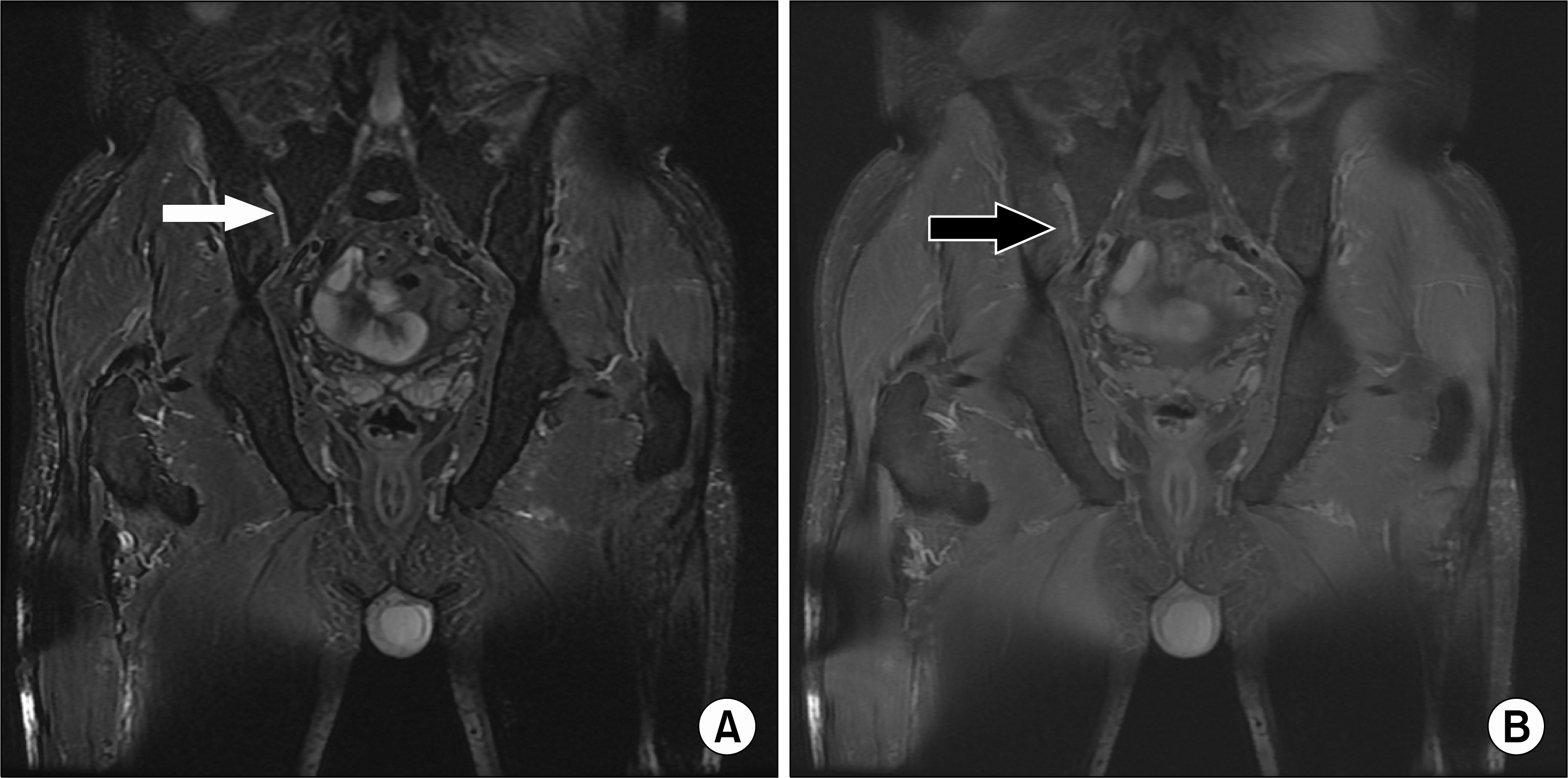 | Figure 1.On pelvic magnetic resonance imaging, subchondral bone edema as an increased signal (white arrow) was noted in the right sacroiliac joint on the fatsaturated fast spin-echo T2-weighted image (A) and coincided with the lesion on the contrast-enhanced fatsaturated fast spin-echo T1 weighted image (black arrow) (B). On the T1 weighted image, there was a fluid collection in the right sacroiliac joint but no abnormal signal was shown around both hip joints. |
 XML Download