Abstract
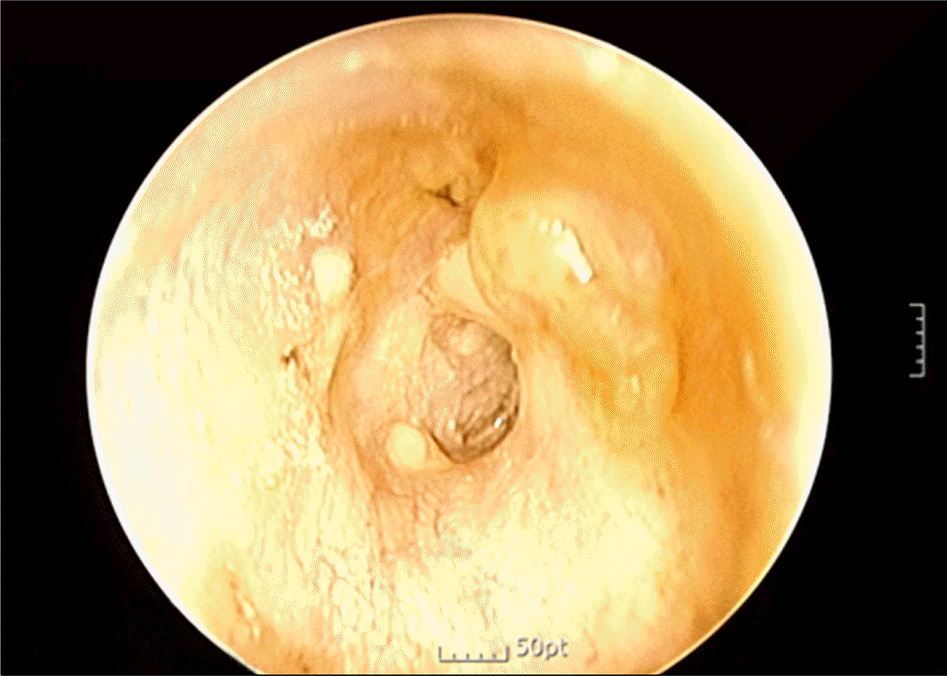
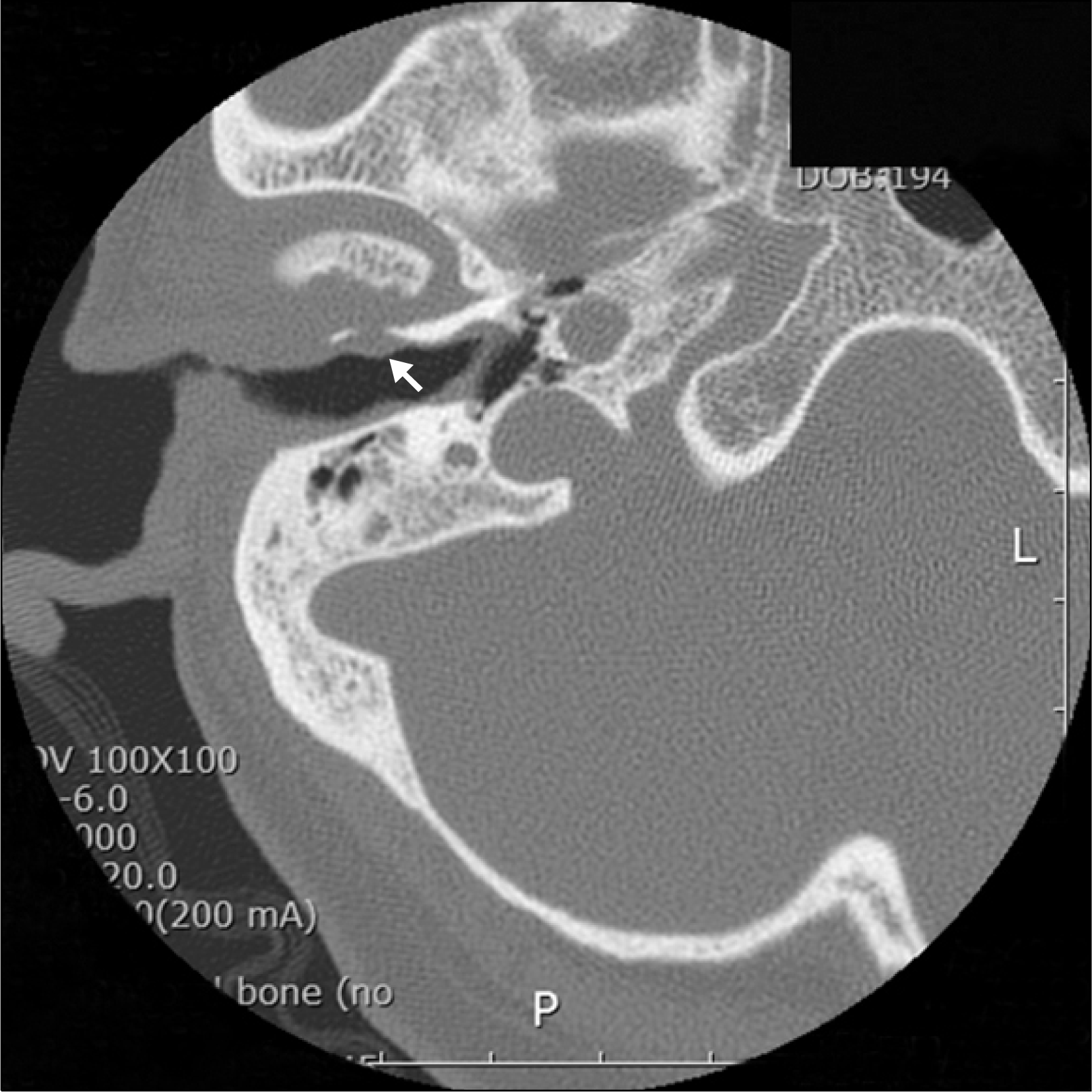
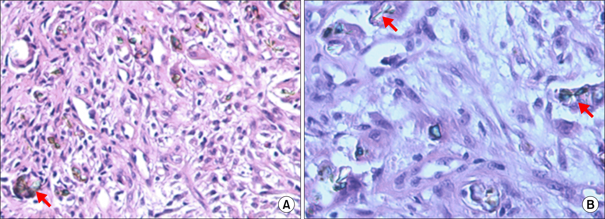
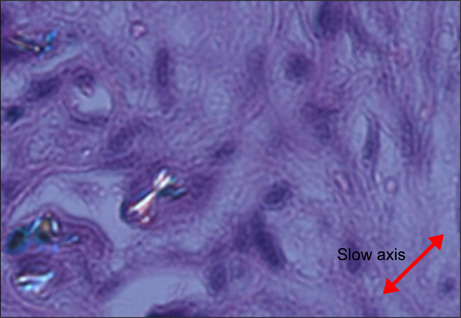
Gout is an inflammatory disorder in which urate crystals are deposited in the joints or soft tissues causing severe inflammation and pain. Urate crystals usually deposit in the joints, and sometimes in the extra-articular sites. A 67-year-old woman visited the otolaryngology clinic due to otalgia and discharge from the ear. She had experienced recurrent arthritis in the left second metatarsophalangeal joint from five years ago. She visited the otolaryngology clinic of our university hospital due to persistent inflammation in the ear canal despite treatment with antibiotics. An otoscopic examination showed a polyp-like mass near the eardrum. Computed tomography scan of temporal bone showed thickening of the eardrum and increased soft tissue density in the external ear canal. On histologic examination the polyp was finally found to be a urate crystal mass. She is now in a good state with urate lowering therapy. We report on a Korean case of tophaceous gout in the external ear canal that was misidentified as an inflammatory polyp.
REFERENCES
1. Reineke U, Ebmeyer J, Schütte F, Upile T, Sudhoff HH. Tophaceous gout of the middle ear. Otol Neurotol. 2009; 30:127–8.

2. Saliba I, Bouthiller A, Desrochers P, Berthlet F, Dufour JJ. Tophaceous gout and pseudogout of the middle ear and the infratemporal fossa: case report and review of the literature. J Otolaryngol. 2003; 32:269–72.

3. Stark TW, Hirokawa RH. Gout and its manifestations in the head and neck. Otolaryngol Clin North Am. 1982; 15:659–64.

4. Forbess LJ, Fields TR. The broad spectrum of urate crystal deposition: unusual presentations of gouty tophi. Semin Arthritis Rheum. 2012; 42:146–54.

5. Antón FM, García Puig J, Ramos T, González P, Ordás J. Sex differences in uric acid metabolism in adults: evidence for a lack of influence of estradiol-17 beta (E2) on the renal handling of urate. Metabolism. 1986; 35:343–8.
6. Puig JG, Michán AD, Jiménez ML, Pérez de Ayala C, Mateos FA, Capitán CF, et al. Female gout. Clinical spectrum and uric acid metabolism. Arch Intern Med. 1991; 151:726–32.

8. Park YB, Park YS, Lee SC, Yoon SJ, Lee SK. Clinical analysis of gouty patients with normouricaemia at diagnosis. Ann Rheum Dis. 2003; 62:90–2.

9. Schlesinger N, Norquist JM, Watson DJ. Serum urate during acute gout. J Rheumatol. 2009; 36:1287–9.

10. Firestein GS BR, Gabriel SE, McInnes IB, O'Dell JR. Kelley' s textbook of rheumatology. 9th ed.p. 1556. Philadelphia: Elsevier Saunders;2013.
Figure 1.
Otoscopic view of right external ear canal showed small polyp on the anterior wall of external ear canal and thickened tympanic membrane.

Figure 2.
Computed tomography of right temporal bone showed soft tissue density in the external auditory canal. Arrow indicates polyp on anterior wall in the external auditory canal.


 PDF
PDF ePub
ePub Citation
 Print
Print


 XML Download