

 PDF
PDF ePub
ePub Citation
 Print
Print


Abstract
Pneumatosis cystoides intestinalis (PCI), the presence of air within the bowel wall, could be complicated with connective tissue disease. PCI associated with dermatomyositis has rarely been reported. Here, we report on a case of PCI that occurred in a 60-year-old female patient with dermatomyositis, presenting with epigastric discomfort. PCI with pneumoperitoneum was detected on abdominal computed tomography but improved by conservative management without surgery. Treatment for secondary PCI is associated with underlying disease. Also, identification of serious complication, such as bowel perforation, necrosis, and peritonitis, requiring surgical intervention is important.
REFERENCES
1. Heng Y, Schuffler MD, Haggitt RC, Rohrmann CA. Pneumatosis intestinalis: a review. Am J Gastroenterol. 1995; 90:1747–58.
2. Kim SY, Cho OK, Koh B, Kim Y, Song SY. Pneumatosis cystoides intestinalis in patients with antinuclear antibody negative systemic lupus erythematosus and dermatomyositis: report of two cases. J Korean Radiol Soc. 2007; 56:361–4.

3. Kim JY, Kwon YH, Lee SJ, Jang SY, Park SY. Pneumatosis cystoides intestinalis and partial abdominal obstruction in a patient with polymyositis. Intest Res. 2011; 9:234–7.
4. Lee SJ, Park JY, Kwon SA, Koh DH, Choi MH, Jang HJ, et al. A case of pneumatosis cystoids intestinalis with polymyositis. Korean J Gastroenterol. 2011; 57:249–52.
5. Selva-O'Callaghan A, Martínez-Costa X, Solans-Laque R, Mauri M, Capdevila JA, Vilardell-Tarrés M. Refractory adult dermatomyositis with pneumatosis cystoides intestinalis treated with infliximab. Rheumatology (Oxford). 2004; 43:1196–7.
6. Saito M, Tanikawa A, Nakasute K, Tanaka M, Nishikawa T. Additive contribution of multiple factors in the development of pneumatosis intestinalis: a case report and review of the literature. Clin Rheumatol. 2007; 26:601–3.
7. Pieterse AS, Leong AS, Rowland R. The mucosal changes and pathogenesis of pneumatosis cystoides intestinalis. Hum Pathol. 1985; 16:683–8.
8. Yale CE, Balish E, Wu JP. The bacterial etiology of pneumatosis cystoides intestinalis. Arch Surg. 1974; 109:89–94.
9. Hall RR, Anagnostou A, Kanojia M, Zander A. Pneumatosis intestinalis associated with graft-versus-host disease of the intestinal tract. Transplant Proc. 1984; 16:1666–8.
10. Jamart J. Pneumatosis cystoides intestinalis. A statistical study of 919 cases. Acta Hepatogastroenterol (Stuttg). 1979; 26:419–22.
11. Park JW, Song YW, Shin KC. A case of pneumatosis cystoides intestinalis in a patient with systemic lupus erythematosus. Korean J Med. 2012; 83:283–6.
12. Knechtle SJ, Davidoff AM, Rice RP. Pneumatosis intestinalis. Surgical management and clinical outcome. Ann Surg. 1990; 212:160–5.
13. Ellis BW. Symptomatic treatment of primary pneumatosis coli with metronidazole. Br Med J. 1980; 280:763–4.
14. Gruenberg JC, Batra SK, Priest RJ. Treatment of pneumatosis cystoides intestinalis with oxygen. Arch Surg. 1977; 112:62–4.
15. Maltz C. Benign pneumoperitoneum and pneumatosis intestinalis. Am J Emerg Med. 2001; 19:242–3.
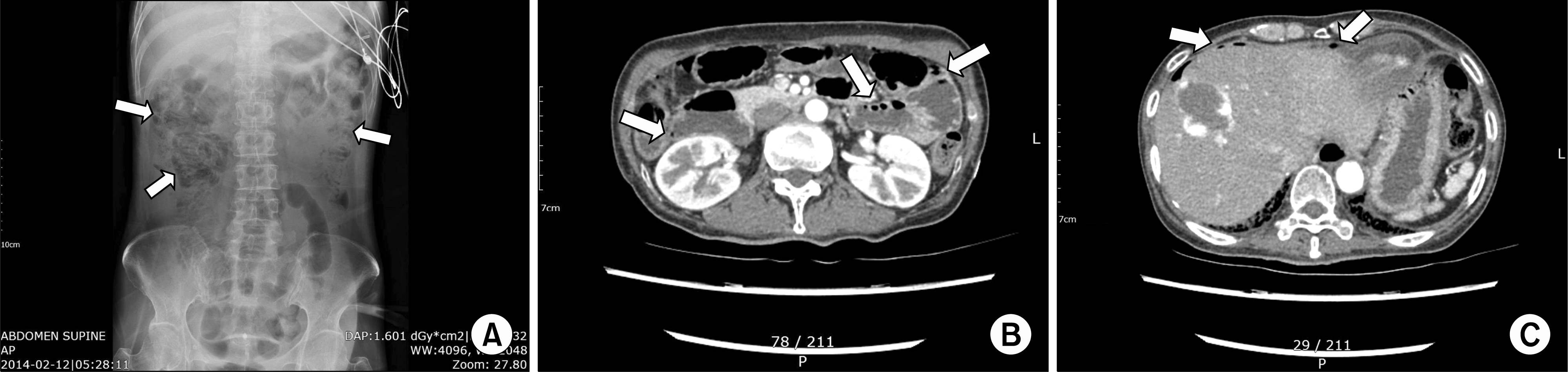
Figure 1.
Abdominal radiography and computed tomography (CT) scan at admission. (A) Abdominal radiography showed linear and cystic air collections in the bowel wall (arrows). (B) Abdominal CT scan revealed several intramural air-filled cysts involving throughout the small bowel (arrows). (C) Multiple small-size free air bubbles were observed in the abdominal cavity, suggesting small bowel micro-perforation (arrows).
 XML Download