

 PDF
PDF ePub
ePub Citation
 Print
Print


Abstract
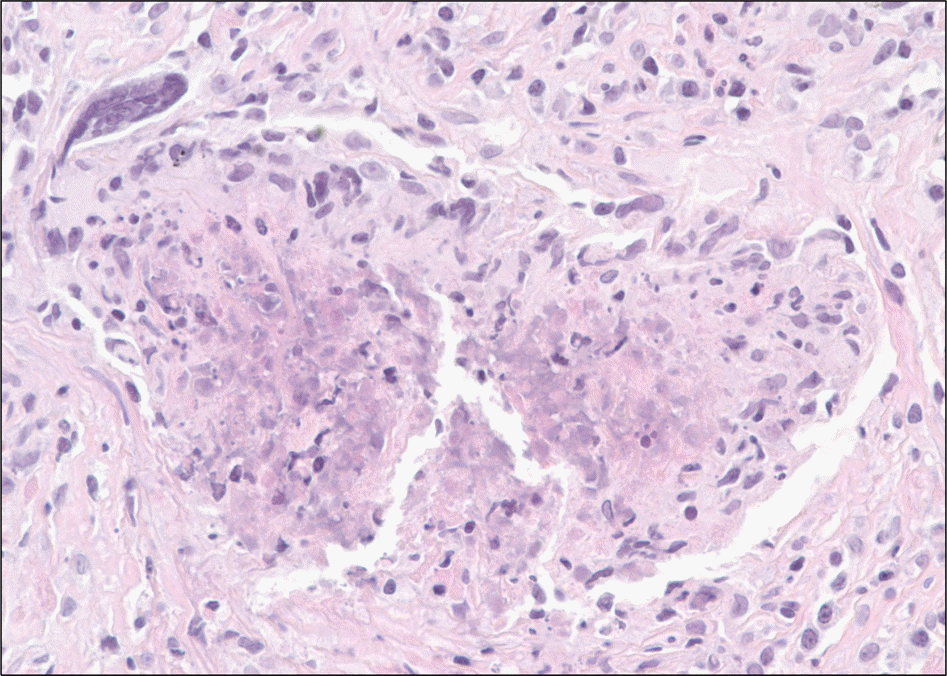
We report on a case of limited form of granulomatosis with polyangiitis (GPA) with pituitary involvement which presented with central diabetes insipidus. This rare form of GPA has not been reported in Korea. The patient presented with fever, headache, productive cough, nasal symptoms, and polyuria. Laboratory data and imaging studies demonstrated inflammatory lesions in nasal sinus and lungs. Pituitary stalk thickening and enhancement were observed on brain magnetic resonance imaging. The histopathology of the lung lesions showed chronic active granulomatous inflammation. Polyuria, hyperosmolar hypernatremia, and decreased urine osmolality which responded to synthetic vasopressin analog were consistent with central diabetes insipidus. Based on the clinical findings and histopathological results, a diagnosis of GPA with pituitary involvement was established. Treatment with desmopressin as well as concurrent glucocorticoids and immunosuppressant resulted in clinical improvement.
Go to : 
REFERENCES
1. Nishino H, Rubino FA, DeRemee RA, Swanson JW, Parisi JE. Neurological involvement in Wegener's granulomatosis: an analysis of 324 consecutive patients at the Mayo Clinic. Ann Neurol. 1993; 33:4–9.

2. Asakura K, Muto T. Neurological involvement in Wegener's granulomatosis. Brain Nerve. 2013; 65:1311–7.
3. Hurst NP, Dunn NA, Chalmers TM. Wegener's granulomatosis complicated by diabetes insipidus. Ann Rheum Dis. 1983; 42:600–1.
4. Garovic VD, Clarke BL, Chilson TS, Specks U. Diabetes insipidus and anterior pituitary insufficiency as presenting features of Wegener's granulomatosis. Am J Kidney Dis. 2001; 37:E5.
5. Düzgün N, Morris Y, Güllü S, Gürsoy A, Ensari A, Kumbasar OO, et al. Diabetes insipidus presentation before renal and pulmonary features in a patient with Wegener's granulomatosis. Rheumatol Int. 2005; 26:80–2.
6. Yong TY, Li JY, Amato L, Mahadevan K, Phillips PJ, Coates PS, et al. Pituitary involvement in Wegener's granulomatosis. Pituitary. 2008; 11:77–84.
7. Barlas NB, Hassan HH, Al Badr FB, Bilal A. Structural and functional involvement of pituitary gland in Wegener's granulomatosis. Clin Neuroradiol. 2011; 21:31–3.
8. Hughes J, Barkhoudarian G, Ciarlini P, Laws ER, Mody E, Inzucchi SE, et al. Refractory pituitary granulomatosis with polyangiitis (Wegener's) treated with rituximab. Endocr Pract. 2013; 19:e1–7.
9. Spísek R, Kolouchová E, Jensovský J, Rusina R, Fendrych P, Plas J, et al. Combined CNS and pituitary involvement as a primary manifestation of Wegener granulomatosis. Clin Rheumatol. 2006; 25:739–42.
10. Hoffman GS, Kerr GS, Leavitt RY, Hallahan CW, Lebovics RS, Travis WD, et al. Wegener granulomatosis: an analysis of 158 patients. Ann Intern Med. 1992; 116:488–98.
11. Xue J, Wang H, Wu H, Jin Q. Wegener's granulomatosis complicated by central diabetes insipidus and peripheral neutrophy with normal pituitary in a patient. Rheumatol Int. 2009; 29:1213–7.
12. Mukhopadhyay S, Gal AA. Granulomatous lung disease: an approach to the differential diagnosis. Arch Pathol Lab Med. 2010; 134:667–90.
13. Hoffman GS, Sechler JM, Gallin JI, Shelhamer JH, Suffredini A, Ognibene FP, et al. Bronchoalveolar lavage analysis in Wegener's granulomatosis. A method to study disease pathogenesis. Am Rev Respir Dis. 1991; 143:401–7.
14. Drent M, Mansour K, Linssen C. Bronchoalveolar lavage in sarcoidosis. Semin Respir Crit Care Med. 2007; 28:486–95.
15. Travis WD, Hoffman GS, Leavitt RY, Pass HI, Fauci AS. Surgical pathology of the lung in Wegener's granulomatosis. Review of 87 open lung biopsies from 67 patients. Am J Surg Pathol. 1991; 15:315–33.
16. Carruthers DM, Connor S, Howie AJ, Exley AR, Raza K, Bacon PA, et al. Percutaneous image-guided biopsy of lung nodules in the assessment of disease activity in Wegener's granulomatosis. Rheumatology (Oxford). 2000; 39:776–82.
17. Leavitt RY, Fauci AS, Bloch DA, Michel BA, Hunder GG, Arend WP, et al. The American College of Rheumatology 1990 criteria for the classification of Wegener's granulomatosis. Arthritis Rheum. 1990; 33:1101–7.
18. Hoffman GS, Leavitt RY, Kerr GS, Fauci AS. The treatment of Wegener's granulomatosis with glucocorticoids and methotrexate. Arthritis Rheum. 1992; 35:1322–9.
19. Bosch X, Guilabert A, Font J. Antineutrophil cytoplasmic antibodies. Lancet. 2006; 368:404–18.
20. Bartůnková J, Tesar V, Sedivá A. Diagnostic and pathogenetic role of antineutrophil cytoplasmic autoantibodies. Clin Immunol. 2003; 106:73–82.
21. Song JG, Lee EJ, Ha ES, Kang EH, Lee SY, In KH, et al. ANCA-negative limited Wegener's granulomatosis. Korean J Med. 2009; 76(Suppl 1):S103–7.
Go to :
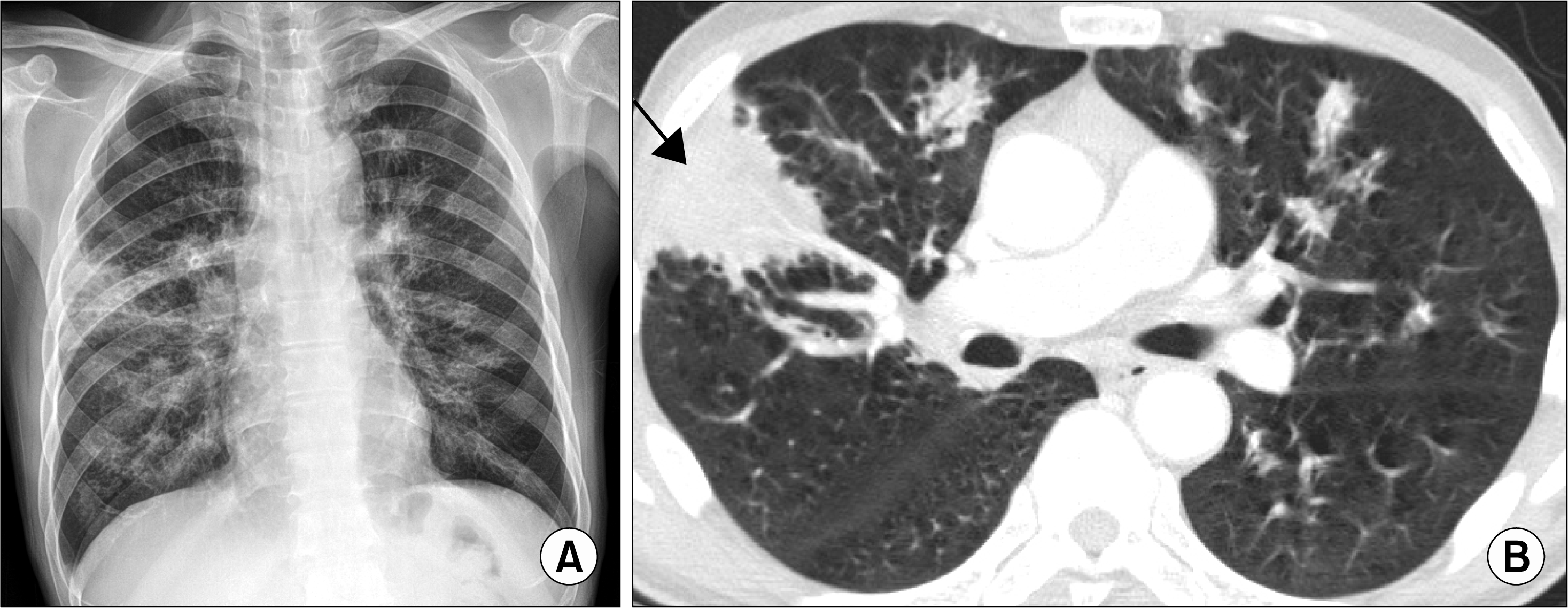 | Figure 1.(A) Chest radiograph shows multifocal patchy consolidations in both lungs. (B) Chest computed tomography demonstrated bilateral infiltrations along the bronchovascular bundles (arrow). |
 | Figure 2.(A, B) Baseline brain magnetic resonance imaging (MRI) of T1 weighted image with contrast showing thickening and enhancement of pituitary stalk (arrows). Leptomeningeal and pa-chymeningeal involvement of left hemisphere are also seen (arrow head). (C, D) Follow-up MRI shows resolustion of pituitary stalk thickening and enhancement (arrows), as well as pachy-meningeal enhancement. |
 XML Download