

 PDF
PDF ePub
ePub Citation
 Print
Print


Abstract
Systemic sclerosis as a connective tissue disease could affect all internal organs of the body and could also manifest as a cutaneous lesion. Cardiac involvement leading to cardiac manifestations in systemic sclerosis patients is not rare. However, cardiac amyloidosis combined with systemic sclerosis is extremely rare. Although there were no definite treatment options in this case, symptomatic treatment is the cornerstone of the management plan. In this case report, we described a correct diagnosis and symptomatic medical care of early reactive cardiac amyloidosis with systemic sclerosis and summarize the current state of the relevant literature.
Go to : 
REFERENCES
1. LeRoy EC, Black C, Fleischmajer R, Jablonska S, Krieg T, Medsger TA Jr, et al. Scleroderma (systemic sclerosis): classification, subsets and pathogenesis. J Rheumatol. 1988; 15:202–5.
2. Kahan A, Coghlan G, McLaughlin V. Cardiac complications of systemic sclerosis. Rheumatology (Oxford). 2009; 48(Suppl 3):iii45–8.

3. Steen VD, Medsger TA Jr. Severe organ involvement in systemic sclerosis with diffuse scleroderma. Arthritis Rheum. 2000; 43:2437–44.
4. Ferri C, Valentini G, Cozzi F, Sebastiani M, Michelassi C, La Montagna G, et al. Systemic Sclerosis Study Group of the Italian Society of Rheumatology (SIR-GSSSc). Systemic sclerosis: demographic, clinical, and serologic features and survival in 1,012 Italian patients. Medicine (Baltimore). 2002; 81:139–53.
5. Kholová I, Kautzner J. Current treatment in cardiac amyloidosis. Curr Treat Options Cardiovasc Med. 2006; 8:468–73.
6. Perfetto F, Moggi-Pignone A, Livi R, Tempestini A, Bergesio F, Matucci-Cerinic M. Systemic amyloidosis: a challenge for the rheumatologist. Nat Rev Rheumatol. 2010; 6:417–29.
7. Scussel-Lonzetti L, Joyal F, Raynauld JP, Roussin A, Rich E, Goulet JR, et al. Predicting mortality in systemic sclerosis: analysis of a cohort of 309 French Canadian patients with emphasis on features at diagnosis as predictive factors for survival. Medicine (Baltimore). 2002; 81:154–67.
8. Black MM. Primary localised cutaneous amyloidosis in systemic sclerosis. Trans St Johns Hosp Dermatol Soc. 1971; 57:177–80.
9. Candell-Riera J, Armadans-Gil L, Simeón CP, Castell-Conesa J, Fonollosa-Pla V, García-del-Castillo H, et al. Comprehensive noninvasive assessment of cardiac involvement in limited systemic sclerosis. Arthritis Rheum. 1996; 39:1138–45.
10. Janosik DL, Osborn TG, Moore TL, Shah DG, Kenney RG, Zuckner J. Heart disease in systemic sclerosis. Semin Arthritis Rheum. 1989; 19:191–200.
11. Hazenberg BP, van Rijswijk MH. Clinical and therapeutic aspects of AA amyloidosis. Baillieres Clin Rheumatol. 1994; 8:661–90.
12. Wada Y, Kobayashi D, Murakami S, Oda M, Hanawa H, Kuroda T, et al. Cardiac AA amyloidosis in a patient with rheumatoid arthritis and systemic sclerosis: the therapeutic potential of biological reagents. Scand J Rheumatol. 2011; 40:402–4.
13. Esplin BL, Gertz MA. Current trends in diagnosis and management of cardiac amyloidosis. Curr Probl Cardiol. 2013; 38:53–96.
14. Lachmann HJ, Goodman HJ, Gilbertson JA, Gallimore JR, Sabin CA, Gillmore JD, et al. Natural history and outcome in systemic AA amyloidosis. N Engl J Med. 2007; 356:2361–71.
Go to :
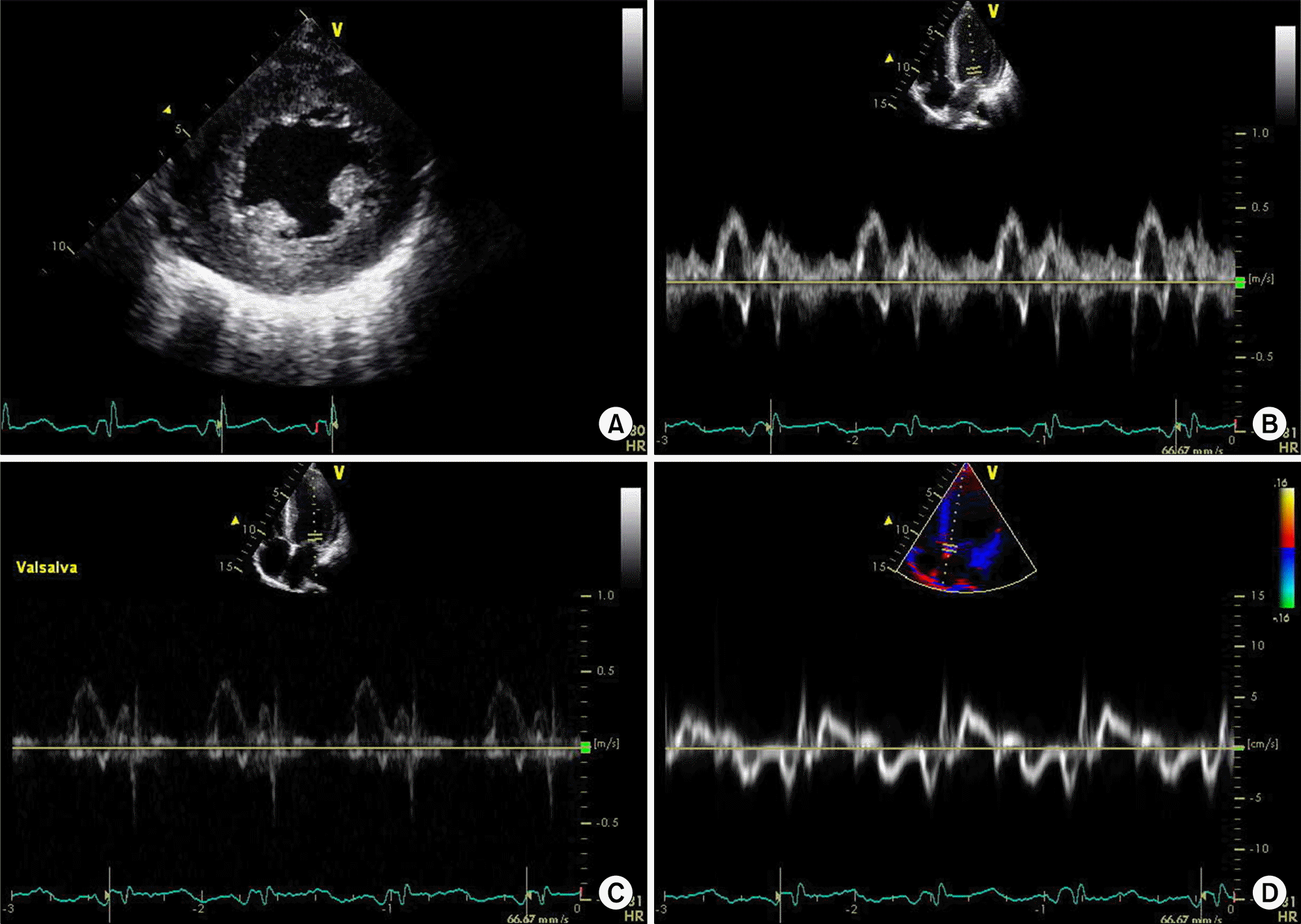 | Figure 1.Echocardiography findings. Echocardiography shows global hypokinesia on left ventricle wall with concentric left ventricle hypertrophy (A), and E wave dominant mitral filling pattern with rapid deceleration time suggests restrictive physiology (B∼ D). |
 XML Download