Abstract
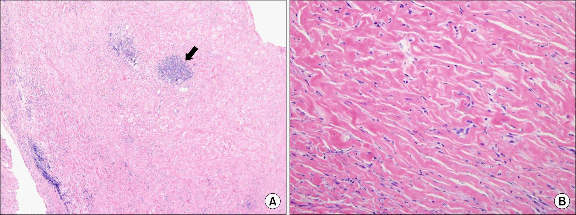
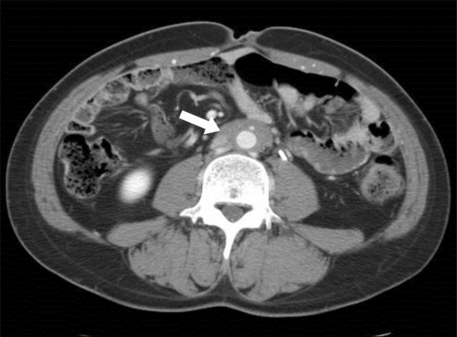
Retroperitoneal fibrosis (RPF) is a rare, progressive disease characterized by chronic non specific inflammation of the retroperitoneum. Although the pathogenesis of idiopathic retroperitoneal fibrosis (IRF) remains unclear, IRF has been reported in association with autoimmune disorders. However, few cases of IRF associated with rheumatoid arthritis (RA) have been reported. We experienced a rare case of IRF in a patient with RA and chronic B viral hepatitis. A 39-year-old Korean man with RA and hepatitis B was referred to our hospital due to left hydronephrosis. An abdominal computed tomography (CT) scan and magnetic resonance imaging (MRI) showed a diffuse infiltrating retroperitoneal mass around the abdominal aorta and left ureter. The patient underwent intraureteral stent insertion and was treated with corticosteroid. Three months later, the follow up abdominal CT showed that the retroperitoneal mass had decreased in size. Herein, we report the first case of coexistent IRF, RA, and chronic B viral hepatitis with a literature review.
REFERENCES
2. Pipitone N, Vaglio A, Salvarani C. Retroperitoneal fibrosis. Best Pract Res Clin Rheumatol. 2012; 26:439–48.

3. Vaglio A, Corradi D, Manenti L, Ferretti S, Garini G, Buzio C. Evidence of autoimmunity in chronic periaortitis: a prospective study. Am J Med. 2003; 114:454–62.

4. Miller OF, Smith LJ, Ferrara EX, McAleer IM, Kaplan GW. Presentation of idiopathic retroperitoneal fibrosis in the pediatric population. J Pediatr Surg. 2003; 38:1685–8.

5. Tsai TC, Chang PY, Chen BF, Huang FY, Shih SL. Retroperitoneal fibrosis and juvenile rheumatoid arthritis. Pediatr Nephrol. 1996; 10:208–9.

6. Couderc M, Mathieu S, Dubost JJ, Soubrier M. Retroperitoneal fibrosis during etanercept therapy for rheumatoid arthritis. J Rheumatol. 2013; 40:1931–3.

7. Vaglio A, Palmisano A, Ferretti S, Alberici F, Casazza I, Salvarani C, et al. Peripheral inflammatory arthritis in patients with chronic periaortitis: report of five cases and review of the literature. Rheumatology (Oxford). 2008; 47:315–8.

8. Corradi D, Maestri R, Palmisano A, Bosio S, Greco P, Manenti L, et al. Idiopathic retroperitoneal fibrosis: clinicopathologic features and differential diagnosis. Kidney Int. 2007; 72:742–53.

9. Martorana D, Vaglio A, Greco P, Zanetti A, Moroni G, Salvarani C, et al. Chronic periaortitis and HLA-DRB1*03: another clue to an autoimmune origin. Arthritis Rheum. 2006; 55:126–30.

10. Rodríguez-Hernández MJ, Viciana P, Cordero E, López-Cortés LF, Pachón J. Retroperitoneal fibrosis in a patient with human immunodeficiency virus infection. Arch Intern Med. 1998; 158:301–2.
11. Hofbauer LC, Magerstadt RA, Heufelder AE. Hepatitis C related cryoglobulinemia associated with retroperitoneal fibrosis. J Rheumatol. 1996; 23:554–7.
12. Pelkmans LG, Aarnoudse AJ, Hendriksz TR, van Bommel EF. Value of acutephase reactants in monitoring disease activity and treatment response in idiopathic retroperitoneal fibrosis. Nephrol Dial Transplant. 2012; 27:2819–25.

13. Vaglio A, Versari A, Fraternali A, Ferrozzi F, Salvarani C, Buzio C. (18)F-fluorodeoxyglucose positron emission tomography in the diagnosis and followup of idiopathic retroperitoneal fibrosis. Arthritis Rheum. 2005; 53:122–5.

Figure 1.
(A) Abdominal computed tomography shows a diffuse infiltrating soft tissue mass (about 5.0×2.5×10.3 cm sized) extending from the lower abdominal aorta to the level of the iliac bifurcation and left ureteral obstruction (arrow). (B) Atrophic change on left kidney (arrowhead).


 PDF
PDF ePub
ePub Citation
 Print
Print


 XML Download