Abstract
Behcet's disease is a systemic inflammatory disorder of unknown etiology, characterized by recurrent oral aphthous ulcers, genital ulcers, uveitis, and skin lesions. Renal involvement is rare in patients with Behcet's disease particularly immunoglobulin A (IgA) nephropathy. Other autoimmune diseases have been associated with increased risk of malignancy, but not Behcet's disease. Some cases of Behcet's disease accompanied by bladder cancer, thyroid cancer, stomach cancer, or hematologic malignancies have been reported. However, to the best of our knowledge, co-occurrence of Behcet's diseases with thymic carcinoma has not yet been reported. We experienced a 49-year-old male patient who had been treated for Behcet disease and IgA nephropathy, who presented with a large mediastinal mass on chest x-ray. After thymectomy, he was diagnosed with thymic carcinoma with complete resection.
REFERENCES
2. Akpolat T, Akkoyunlu M, Akpolat I, Dilek M, Odabas AR, Ozen S. Renal Behçet's disease: a cumulative analysis. Semin Arthritis Rheum. 2002; 31:317–37.

3. Altay M, Secilmis S, Unverdi S, Ceri M, Duranay M. Behcet's disease and IgA nephropathy. Rheumatol Int. 2012; 32:2227–9.

4. Ahn JK, Oh JM, Lee J, Koh EM, Cha HS. Behcet's disease associated with malignancy in Korea: a single center experience. Rheumatol Int. 2010; 30:831–5.

5. Hill CL, Zhang Y, Sigurgeirsson B, Pukkala E, Mellemkjaer L, Airio A, et al. Frequency of specific cancer types in dermatomyositis and polymyositis: a population-based study. Lancet. 2001; 357:96–100.

6. Kaklamani VG, Tzonou A, Kaklamanis PG. Behçet's disease associated with malignancies. Report of two cases and review of the literature. Clin Exp Rheumatol. 2005; 23(4 Suppl 38):S35–41.
7. Cengiz M, Altundag MK, Zorlu AF, Güllü IH, Ozyar E, Atahan IL. Malignancy in Behçet's disease: a report of 13 cases and a review of the literature. Clin Rheumatol. 2001; 20:239–44.

8. Kural-Seyahi E, Fresko I, Seyahi N, Ozyazgan Y, Mat C, Hamuryudan V, et al. The long-term mortality and morbidity of Behçet syndrome: a 2-decade outcome survey of 387 patients followed at a dedicated center. Medicine (Baltimore). 2003; 82:60–76.
9. Roden AC, Yi ES, Cassivi SD, Jenkins SM, Garces YI, Aubry MC. Clinicopathological features of thymic carcinomas and the impact of histopathological agreement on prognostical studies. Eur J Cardiothorac Surg. 2013; 43:1131–9.

11. Okereke IC, Kesler KA, Freeman RK, Rieger KM, Birdas TJ, Ascioti AJ, et al. Thymic carcinoma: outcomes after surgical resection. Ann Thorac Surg. 2012; 93:1668–72. discussion 1672-3.

13. Tamaoki N, Habu S, Yoshimatsu H, Tsuchiya M, Watanabe H. Thymic change in Behçet's disease. Keio J Med. 1972; 21:201–13.

14. Posner MR, Prout MN, Berk S. Thymoma and the nephrotic syndrome: a report of a case. Cancer. 1980; 45:387–91.

15. Schillinger F, Milcent T, Wolf C, Gulino R, Montagnac R. Nephrotic syndrome revealing malignant thymoma. Presse Med. 1998; 27:60–3.
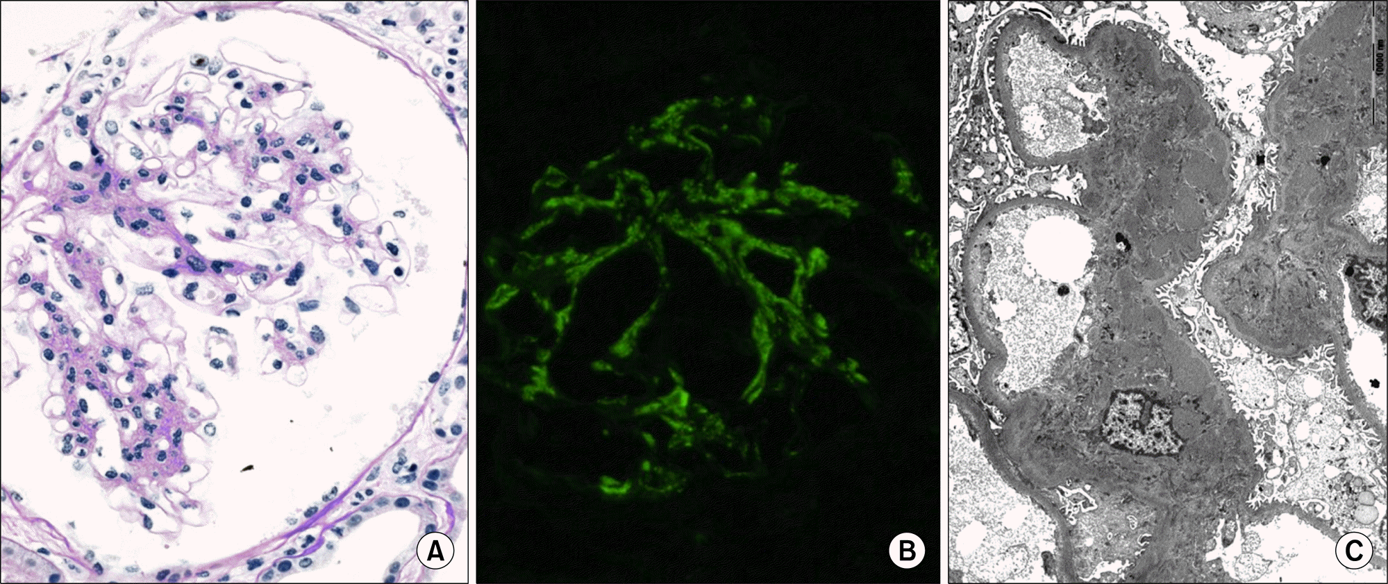
Figure 1.
(A) On light microscopic finding, mesangial cell proliferaion and mesangial matrix expansion are observed (periodic acid-Schiff stain, ×400). (B) On immunofluorescent study, immnofluorescent activity for immunoglobulin A is obsereved on the mesangium (×400). (C) Electron microscopic examination reveals electron dense deposits on the mesangium and paramesangium (×4,500).

Figure 2.
(A) A chest posterial-anterial (PA) shows a large right anterior mediastinal mass (arrows) which does not shown on 18 months ago chest PA (B). (C) A chest computed tomography demonstrates a large right anterior mediastinal mass (arrows) which shows mixed solid and cystic components.

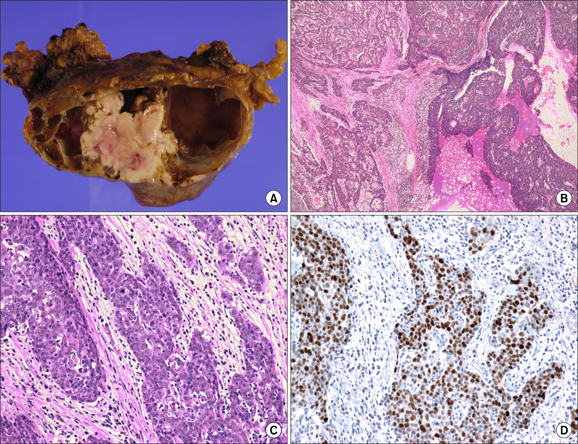
Figure 3.
(A) On gross examination, the mass is relatively well demarcated and on cut section, it shows multilocular cysts containing clear and mucoid fluid and partly whitish solid areas with necrosis. (B) On lower power microscopic examination, the cystic area corresponds to the thymoma and the solid area corresponds to the thymic carcinoma showing infiltrative pattern (H&E, ×40).(C) On high power field, the thymic carcinoma area shows histology of squamous cell carcinoma (H&E, ×200). (D) On immunohistochemical stain, these tumor cells are positive for p53 (×200).


 PDF
PDF ePub
ePub Citation
 Print
Print


 XML Download