Abstract
Rheumatoid arthritis (RA) is a systemic autoimmune disorder which can affect all of the synovial joints including the cervical spine. Cervical involvement typically begins early in the disease process and shows relatively slow progression. Fractures of the odontoid process are mainly noted after a major trauma to the cervical spine. A case of a 77-year-old woman with paresthesia of the extremities caused by spontaneous atraumatic fracture of the odontoid process, which was revealed as a manifestation of RA, is presented in this report.
Go to : 
REFERENCES
1. da Côrte FC, Neves N. Cervical spine instability in rheumatoid arthritis. Eur J Orthop Surg Traumatol. 2014; 24(Suppl 1):S83–91.

2. Al Khayer A, Sawant N, Emberton P, Sell PJ. Spontaneous odontoid process fracture in rheumatoid arthritis: diagnostic difficulties, pathology and treatment. Injury. 2006; 37:659–62.

3. Lewandrowski KU, Park PP, Baron JM, Curtin SL. Atraumatic odontoid fractures in patients with rheumatoid arthritis. Spine J. 2006; 6:529–33.

4. Ballard WT, Clark CR. Increased atlantoaxial instability secondary to an atraumatic fracture of the odontoid process in a patient who had rheumatoid arthritis. A case report. J Bone Joint Surg Am. 1995; 77:1245–8.

5. Toyama Y, Hirabayashi K, Fujimura Y, Satomi K. Spontaneous fracture of the odontoid process in rheumatoid arthritis. Spine (Phila Pa 1976). 1992; 17(10 Suppl):S436–41.

6. Byrne RP, Woodward HW. Occult fracture of the odontoid process: report of case. J Oral Surg. 1972; 30:684–6.
7. Storms GE, Kruijsen MW, Van Beusekom HJ, Van de Putte LB, Boerbooms AM, Penn WH, et al. Pathological fracture of the odontoid process in rheumatoid arthritis. Neth J Med. 1980; 23:120–22.
8. Kim TH, Kim DY, Jun JB, Jung SS, Lee IH, Bae SC, et al. A case of odontoid fracture in rheumatoid arthritis. J Korean Rheum Assoc. 1995; 2:197–201.
9. Aletaha D, Neogi T, Silman AJ, Funovits J, Felson DT, Bingham CO 3rd, et al. 2010 Rheumatoid arthritis classification criteria: an American College of Rheumatology/ European League Against Rheumatism collaborative initiative. Arthritis Rheum. 2010; 62:2569–81.
10. Yurube T, Sumi M, Nishida K, Miyamoto H, Kohyama K, Matsubara T, et al. Kobe Spine Conference. Incidence and aggravation of cervical spine instabilities in rheumatoid arthritis: a prospective minimum 5-year follow-up study of patients initially without cervical involvement. Spine (Phila Pa 1976). 2012; 37:2136–44.
11. Ea HK, Weber AJ, Yon F, Lioté F. Osteoporotic fracture of the dens revealed by cervical manipulation. Joint Bone Spine. 2004; 71:246–50.

12. Elgafy H, Dvorak MF, Vaccaro AR, Ebraheim N. Treatment of displaced type II odontoid fractures in elderly patients. Am J Orthop (Belle Mead NJ). 2009; 38:410–6.
Go to : 
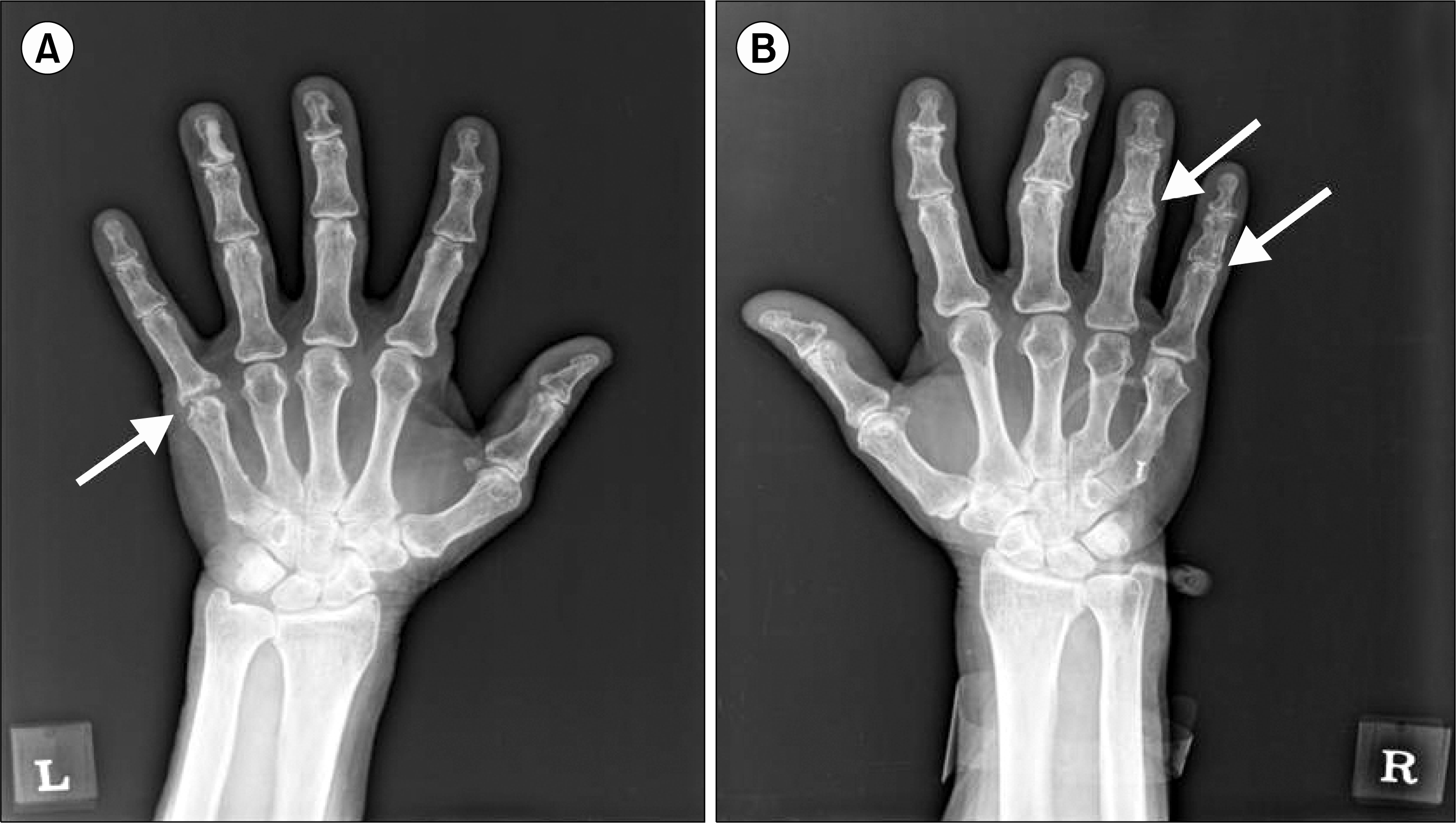
 | Figure 1.Plain radiographs of both hands showed a marginal bone erosion in the left 5th metacarpophalangeal joint (A; white arrow) and joint space narrowing with marginal bone erosion in the right 4th and 5th proximal interphalangeal joints (B; white arrows). |
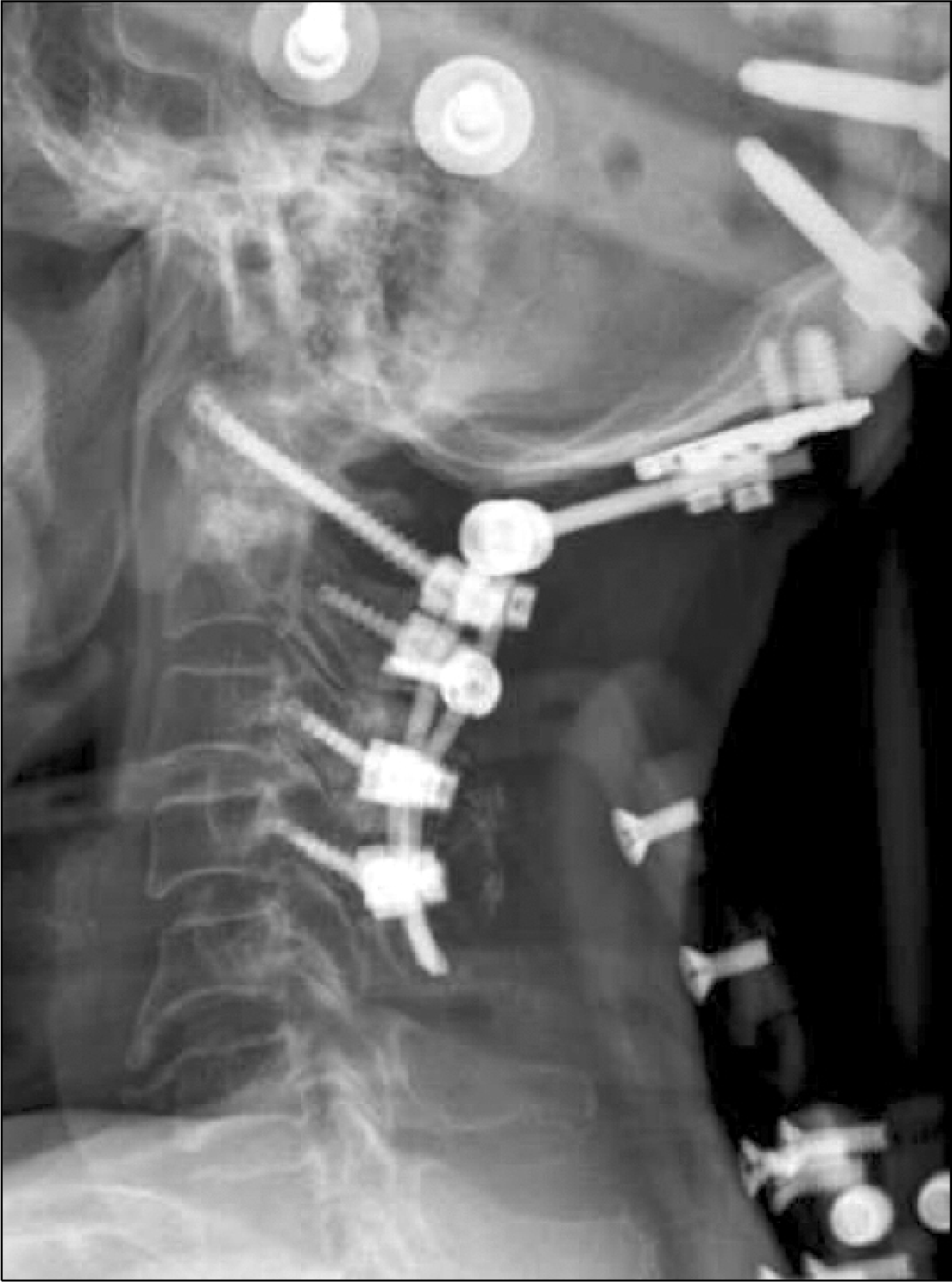
 | Figure 2.Lateral radiographs of the cervical spine in flexion (A) and extension (B). In flexion, the distance between the posterior aspect of the anterior ring of atlas and the anterior surface of the odontoid process (arrowheads) was 6 mm and showed atlantoaxial subluxation. Computed tomography scanning revealed a fracture of the odontoid process (C and D; white arrows). T2 weighted sagittal magnetic resonance imaging revealed C2 level spinal cord compression (E; white arrow). |


 PDF
PDF ePub
ePub Citation
 Print
Print


 XML Download