Abstract
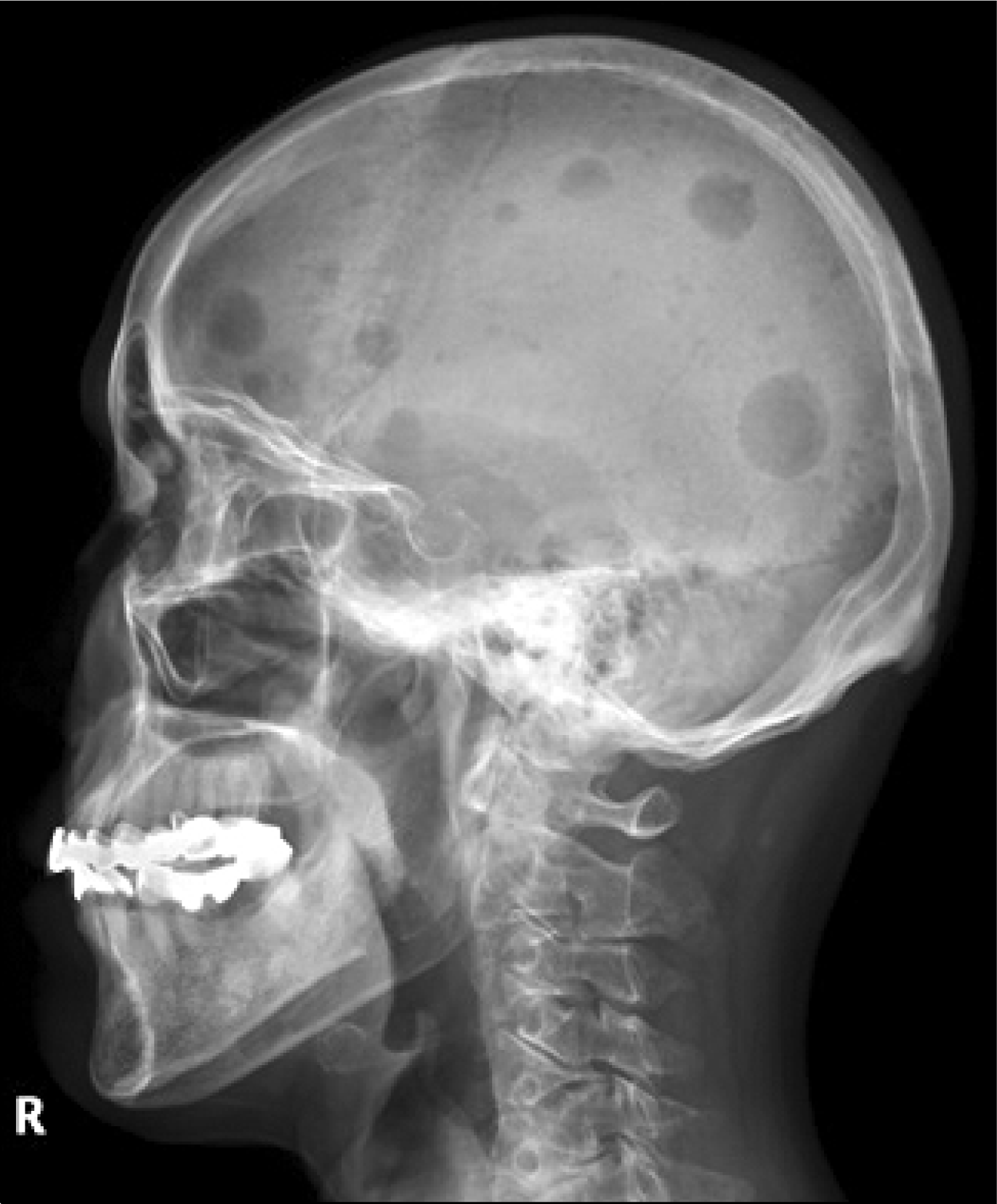
It is known that rheumatoid arthritis (RA) patients show increased incidence of multiple myeloma (MM), despite its rarity. Only one case of MM with seronegative RA was reported in Korea, thus far. We report a case of MM with seropositive RA. The patient was a 66 year old female who had been diagnosed with seropositive RA 4 years ago. Over the last 1 month, the patient experienced general weakness and weight loss of 10 kg. It was found that her serum creatinine had increased and her urine analysis showed proteinuria. To evaluate renal failure and proteinuria, renal biopsy, bone marrow biopsy and electrophoresis were carried out. A diagnosis of myeloma cast nephropathy was made. We report this rare case of MM represented as acute renal failure during the treatment for RA, and include a review of the literature.
REFERENCES
1. Son KM, Kim JK, Kim HA, Park HR, Park EJ, Oh JM, et al. Vasculitis as a presenting feature of multiple myeloma in a patient with rheumatoid arthritis. J Rheum Dis. 2013; 20:44–7.

2. Lee SJ, Lee SS, Kim YA, Park MJ, Lee JJ, Kim HJ. A case of multiple myeloma in a patient with systemic lupus erythematosus. J Korean Rheum Assoc. 2002; 9:325–9.
3. Jorgensen C, Guerin B, Ferrazzi V, Bologna C, Sany J. Arthritis associated with monoclonal gammapathy: clinical characteristics. Br J Rheumatol. 1996; 35:241–3.

4. Cibere J, Sibley J, Haga M. Rheumatoid arthritis and the risk of malignancy. Arthritis Rheum. 1997; 40:1580–6.

5. Eriksson M. Rheumatoid arthritis as a risk factor for multiple myeloma: a case-control study. Eur J Cancer. 1993; 29A:259–63.

6. Kelly C, Sykes H. Rheumatoid arthritis, malignancy, and paraproteins. Ann Rheum Dis. 1990; 49:657–9.

7. Usnarska-Zubkiewicz L, Czarnecka M, Włodarczyk S, Nowak E. Rheumatoid arthritis as a risk factor for development of multiple myeloma. Pol Arch Med Wewn. 1997; 97:253–9.
8. Youinou P, Le Corre R, Dueymes M. Autoimmune diseases and monoclonal gammopathies. Clin Exp Rheumatol. 1996; 14(Suppl 14):S55–8.
9. Pers JO, Jamin C, Predine-Hug F, Lydyard P, Youinou P. The role of CD5-expressing B cells in health and disease (review). Int J Mol Med. 1999; 3:239–45.

10. Matteson EL, Hickey AR, Maguire L, Tilson HH, Urowitz MB. Occurrence of neoplasia in patients with rheumatoid arthritis enrolled in a DMARD Registry. Rheumatoid Arthritis Azathioprine Registry Steering Committee. J Rheumatol. 1991; 18:809–14.
11. Flipo RM, Deprez X, Fardellone P, Duquesnoy B, Delcambre B. Rheumatoid arthritis and multiple myeloma. Apropos of 22 cases. Results of a multicenter national survey. Rev Rhum Ed Fr. 1993; 60:269–73.
12. Wolfe F, Michaud K. Biologic treatment of rheumatoid arthritis and the risk of malignancy: analyses from a large US observational study. Arthritis Rheum. 2007; 56:2886–95.

13. Ardalan MR, Shoja MM. Multiple myeloma presented as acute interstitial nephritis and rheumatoid arthritis-like polyarthritis. Am J Hematol. 2007; 82:309–13.

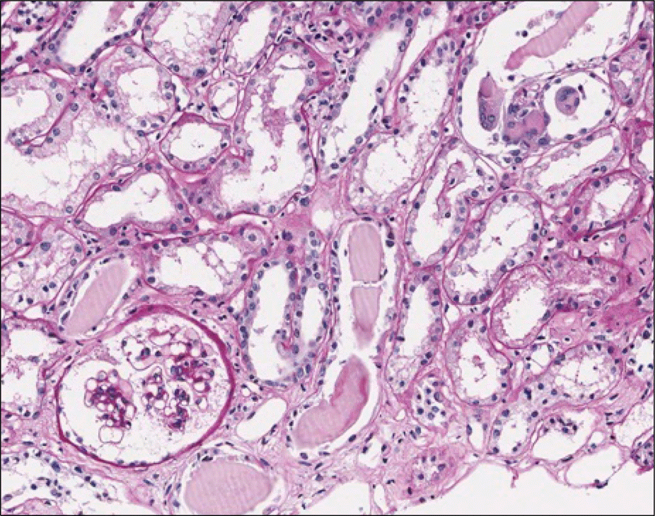
Figure 2.
Renal biopsy shows cast nephropathy. Tubular casts, PAS negative, broken or fractured, are present. Some casts are surrounded by multinucleated giant cells. Glomerulus looks normal (×400).


 PDF
PDF ePub
ePub Citation
 Print
Print


 XML Download