Abstract
Amyloidosis is a clinical disorder caused by extracellular deposition of proteinaceous insoluble fibrils in various tissues, resulting in organ compromise. Amyloid L (AL) amyloidosis is the most common type of systemic amyloidosis, which occurs in association with multiple myeloma or monoclonal gammopathy of undetermined significance (MGUS). Secondary amyloid A (AA) amyloidosis is a complication of chronic inflammatory conditions, such as rheumatoid arthritis or ankylosing spondylitis. We report a case of a 49-year-old manwith a 11-year history of ankylosing spondylitis, who was recently diagnosed with MGUS presented with cardiac amyloidosis of both the AA and AL types. We report this case along with a review of relevant literature.
Go to : 
REFERENCES
1. Cunnane G. Amyloid precursors and amyloidosis in inflammatory arthritis. Curr Opin Rheumatol. 2001; 13:67–73.

3. Desai HV, Aronow WS, Peterson SJ, Frishman WH. Cardiac amyloidosis: approaches to diagnosis and management. Cardiol Rev. 2010; 18:1–11.
4. Wright JR, Calkins E, Humphrey RL. Potassium perman-ganate reaction in amyloidosis. A histologic method to assist in differentiating forms of this disease. Lab Invest. 1977; 36:274–81.
5. Marhaug G, Dowton SB. Serum amyloid A: an acute phase apolipoprotein and precursor of AA amyloid. Baillieres Clin Rheumatol. 1994; 8:553–73.

6. Jung SY, Park MC, Park YB, Lee SK. Serum amyloid a as a useful indicator of disease activity in patients with ankylosing spondylitis. Yonsei Med J. 2007; 48:218–24.

7. Carroll JD, Gaasch WH, McAdam KP. Amyloid cardiomyopathy: characterization by a distinctive volt-age/mass relation. Am J Cardiol. 1982; 49:9–13.

Go to : 
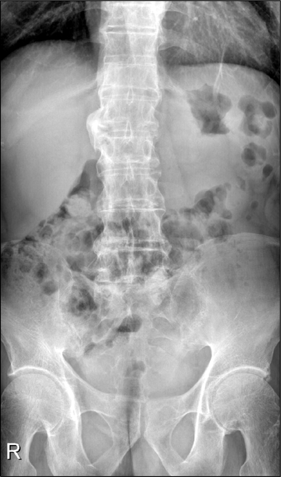 | Figure 1.Lumbar radiography shows squaring of the body with the syndesmophyte along the disc margin of the lumbar spine. Facet anklyosis of the lower lumbar spine. |
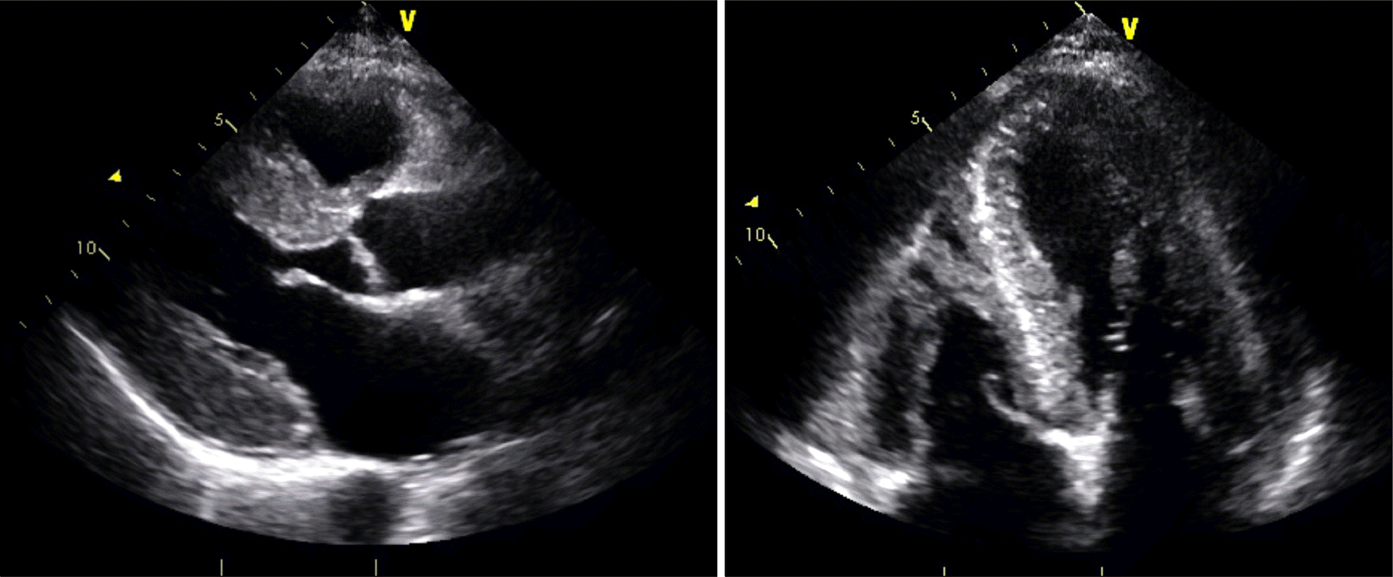 | Figure 2.Echocardiography demonstrated a ground glass opacity pattern of the myocardium and increased wall thickness (septal thickness, 15.9 mm; posterior wall thickness, 14.4 mm). |
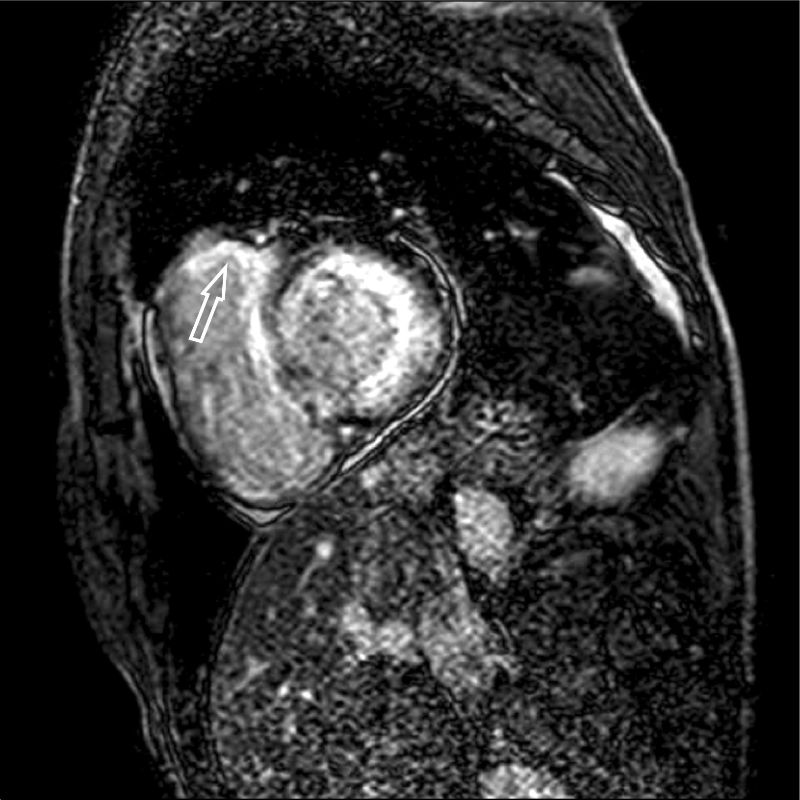 | Figure 3.Midventricular short-axis LGE-CMR image shows global subendocardial LGE of the left ventricle and patchy transmural enhancement of right ventricle (arrow). LGE: late gadolinium enhancement, CMR: cardiac magnetic resonance. |


 PDF
PDF ePub
ePub Citation
 Print
Print


 XML Download