

 PDF
PDF ePub
ePub Citation
 Print
Print


Abstract
In patients with dermatomyositis, chronic inflammation of the pharynx and esophagus results in coughing and diffi-culty in swallowing. These become important clinical symptoms, especially if they contribute to malnutrition or aspiration pneumonia. They can ultimately reduce the quality of life. In rare cases, if the symptoms worsen despite proper treatment, serious complications may arise, a reason to suspect an esophageal perforation or abscess. The authors report a case of dermatomyositis in an adult patient with rare complications of spontaneous esophageal perforation and hypopharyngeal abscess. The patient received non-surgical treatment and was able to resume oral intake of food.
References
1. Iking-Konert C, Ostendorf B, Jung G, Becker A, Schneider M. "Bubbles in the brain": an unusual complication of dermatomyositis. Ann Rheum Dis. 2006; 65:550–1.

2. Ebert EC. Review article: the gastrointestinal complications of myositis. Aliment Pharmacol Ther. 2010; 31:359–65.
3. Williams RB, Grehan MJ, Hersch M, Andre J, Cook IJ. Biomechanics, diagnosis, and treatment outcome in inflammatory myopathy presenting as oropharyngeal dysphagia. Gut. 2003; 52:471–8.
4. Oh TH, Brumfield KA, Hoskin TL, Stolp KA, Murray JA, Bassford JR. Dysphagia in inflammatory myopathy: clinical characteristics, treatment strategies, and outcome in 62 patients. Mayo Clin Proc. 2007; 82:441–7.
5. Du Y, Dai N, Yu H, Lu Z. Tracheoesophageal fistula: a rare complication of adult dermatomyositis. Eur J Dermatol. 2008; 18:347–8.
6. Dougenis D, Papathanasopoulos PG, Paschalis C, Papapet-ropoulos T. Spontaneous esophageal rupture in adult dermatomyositis. Eur J Cardiothorac Surg. 1996; 10:1021–3.
7. Ghandour Z, al Karawi MA, Mohamed AE. Spontaneous esophageal perforation: unusual presentation of tuber-culosis. Endoscopy. 1997; 29:143–4.
8. Al-Shawwa B, D'Andrea L, Quintero D. Candida esophageal perforation and esophagopleural fistula: a case report. J Med Case Rep. 2008; 2:209.
9. Wu JT, Mattox KL, Wall MJ Jr. Esophageal perforations: new perspectives and treatment paradigms. J Trauma. 2007; 63:1173–84.
Figure 1.
(A) Histopathologic features of an incisional biopsy of the vastus lateralis muscle revealed variation in the fiber sizes and focal perifascicular atrophy, ×40. (B) Muscle fibers showing degene-rating and regenerating changes and perivascular infiltration of lymphohistiocytes, ×200.
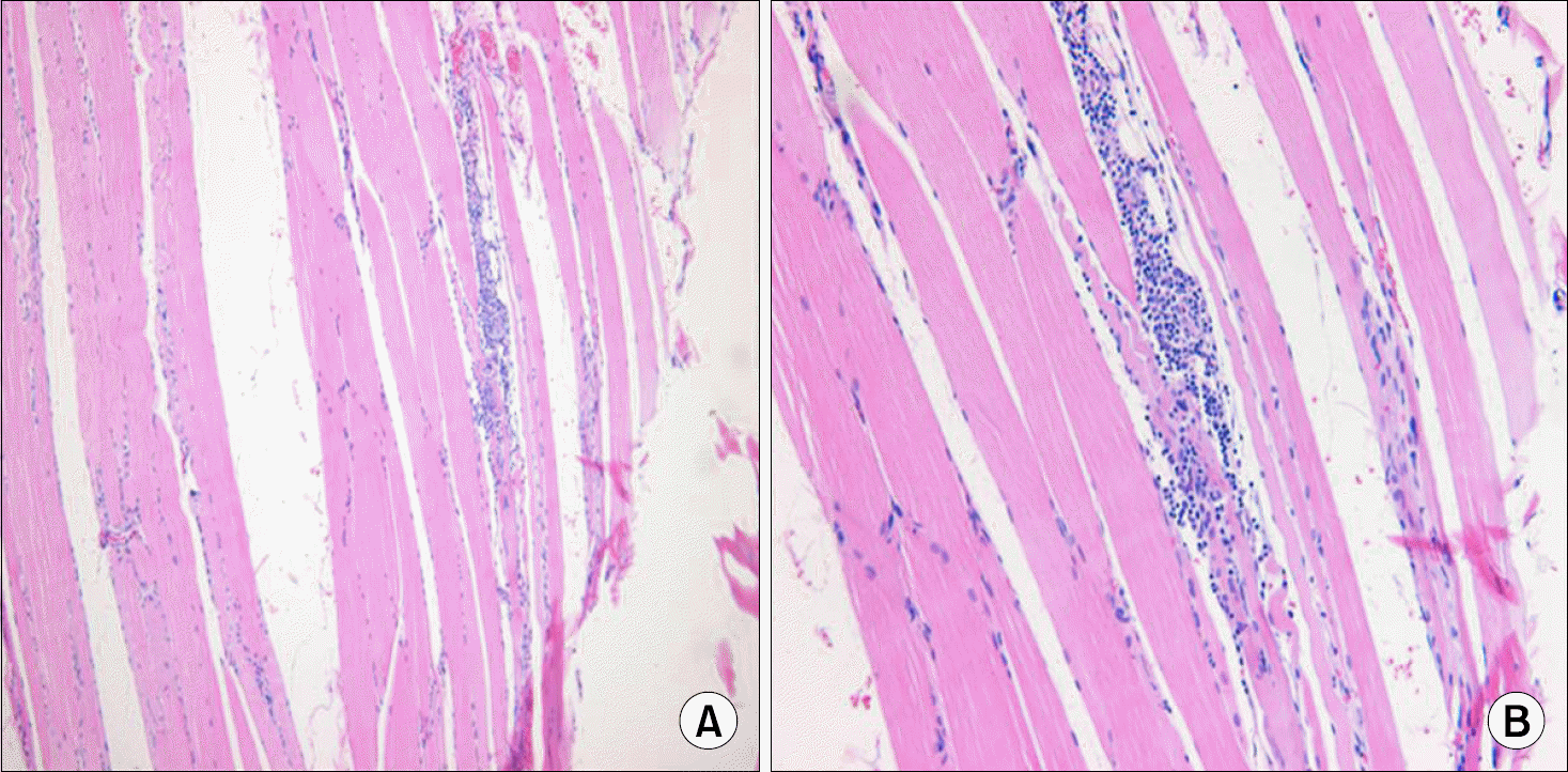
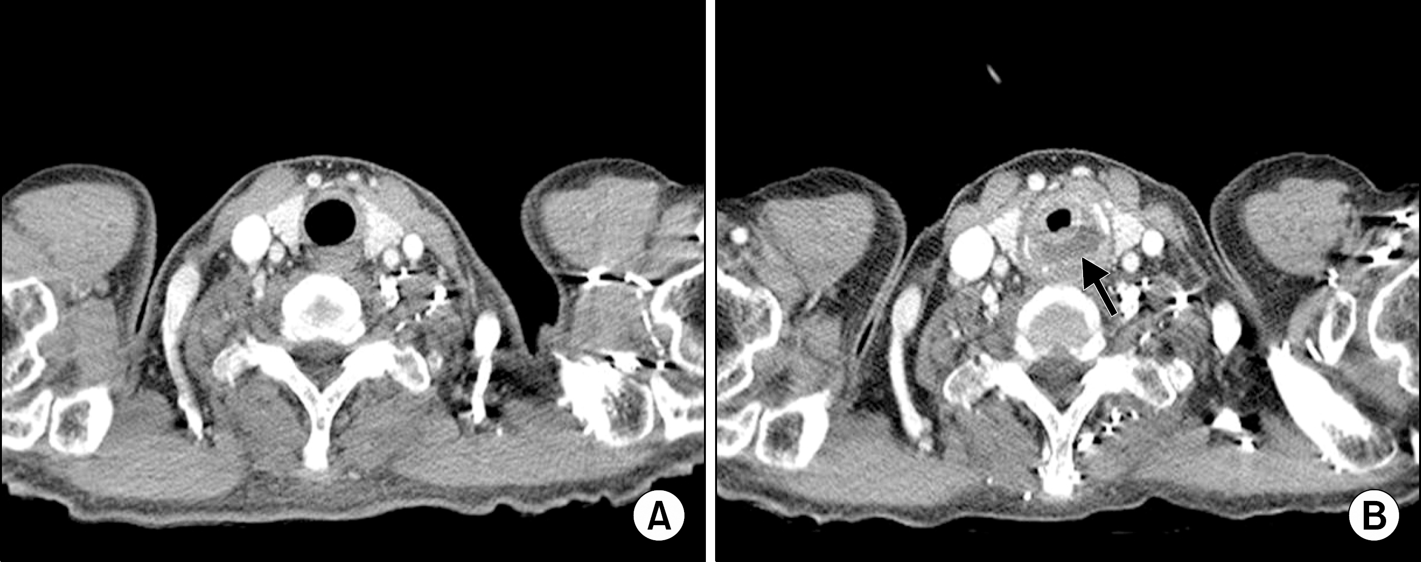
Figure 2.
(A) Computed tomography of the chest showed no remarkable findings in the pharynx in July, 2011. (B) Computed tomography of the chest showed peri-pheral enhancing fluid collection in the inferior aspect of cricoids cartilage, suggesting the presence of an abscess formation in Decem-ber, 2011 (black arrow).
 XML Download