

 PDF
PDF ePub
ePub Citation
 Print
Print


Abstract
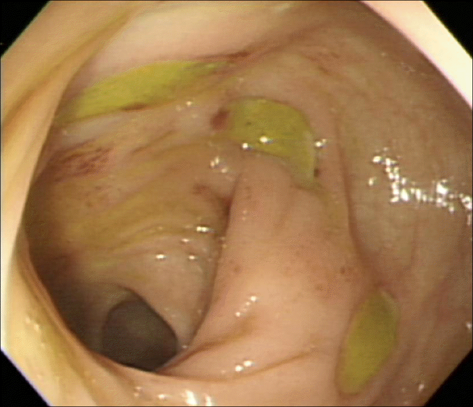
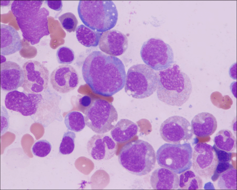
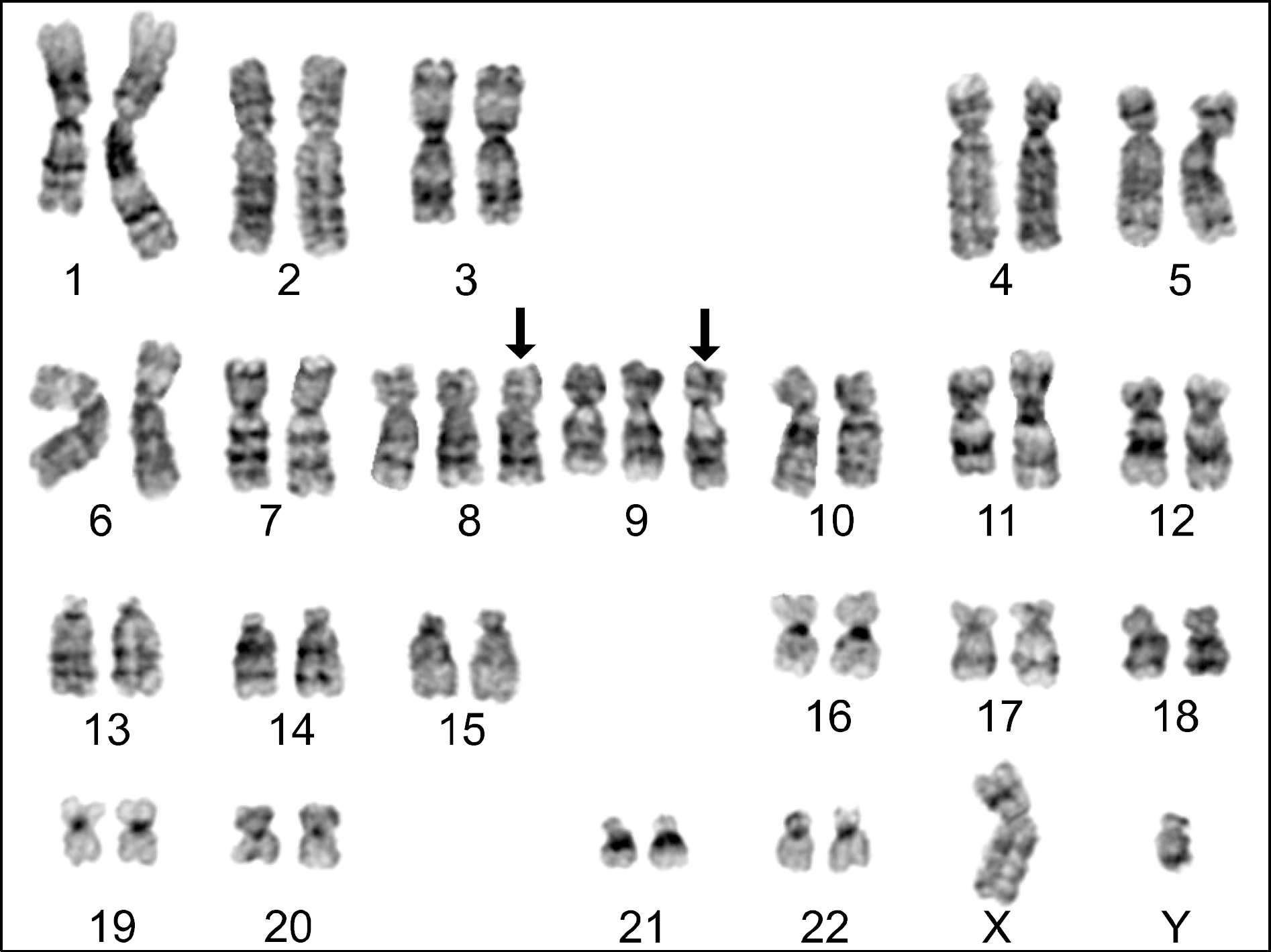
Behcet's disease (BD) is a multi-systemic inflammatory disease of unknown origin that affects nearly all organs. Recent reports of BD with myelodysplastic syndrome (MDS) often note an association with gastrointestinal involvement and trisomy 8. We herein report on a case of a 51-year-old man who had refractory schizophrenia and developed gastrointestinal BD and MDS with trisomy 8 and 9. He visited our hospital due to fever and abdominal pain. Multiple ulcerations in the colorectum were observed on colonoscopy, and he was diagnosed with intestinal BD. During the treatment of intestinal BD, anemia and throm-bocytopenia developed. His bone marrow study revealed myelodysplastic syndrome (refractory anemia with ringed sideroblast) with trisomy 8 and trisomy 9. We report a rare case of intestinal BD accompanied by schizophrenia and myelodysplastic syndrome with trisomy 8 and 9.
Go to : 
References
1. Sakane T, Takeno M, Suzuki N, Inaba G. Behç et's disease. N Engl J Med. 1999; 341:1284–91.
2. Ahn JK, Cha HS, Koh EM, Kim SH, Kim YG, Lee CK, et al. Behcet's disease associated with bone marrow failure in Korean patients: clinical characteristics and the association of intestinal ulceration and trisomy 8. Rheumatology (Oxford). 2008; 47:1228–30.

3. Solé F, Woessner S, Florensa L, Pérez-Losada A, Acín P, Besses C, et al. Cytogenetic study of 93 myelodysplastic syndromes. Med Clin (Barc). 1998; 110:94–8.
4. Chen HC, Chiu YM. Large-vessel thrombosis in intestinal Behç et's disease complicated with myelodysplastic syndrome and trisomy 8. World J Gastroenterol. 2012; 18:1137–40.
5. Ebert EC. Gastrointestinal manifestations of Behç et's disease. Dig Dis Sci. 2009; 54:201–7.
6. Turan B, Gallati H, Erdi H, Gürler A, Michel BA, Villiger PM. Systemic levels of the T cell regulatory cytokines IL-10 and IL-12 in Bechç et's disease; soluble TNFR-75 as a biological marker of disease activity. J Rheumatol. 1997; 24:128–32.
7. Stasi R, Amadori S, Newland AC, Provan D. Infliximab chimeric antitumor necrosis factor-a monoclonal antibody as potential treatment for myelodysplastic syndromes. Leuk Lymphoma. 2005; 46:509–16.
8. Solé F, Prieto F, Badia L, Woessner S, Florensa L, Caballin MR, et al. Cytogenetic studies in 112 cases of untreated myelodysplastic syndromes. Cancer Genet Cytogenet. 1992; 64:12–20.
9. Chen G, Zeng W, Miyazato A, Billings E, Maciejewski JP, Kajigaya S, et al. Distinctive gene expression profiles of CD34 cells from patients with myelodysplastic syndrome characterized by specific chromosomal abnormali-ties. Blood. 2004; 104:4210–8.
10. Watanabe M, Ueno Y, Yajima T, Okamoto S, Hayashi T, Yamazaki M, et al. Interleukin 7 transgenic mice develop chronic colitis with decreased interleukin 7 protein accumulation in the colonic mucosa. J Exp Med. 1998; 187:389–402.
11. Deniz O, Cayköylü A, Vural G, Albayrak Y, Temel S, Aydin I, et al. A case study of neuro-psycho-Behç et's syndrome presenting with psychotic attack. Clin Neurol Neurosurg. 2009; 111:877–9.
12. Sperber MA. Schizophrenia and organic brain syndrome with trisomy 8 (group-C trisomy 8 [47, XX, 8+]). Biol Psychiatry. 1975; 10:27–43.
13. Blouin JL, Dombroski BA, Nath SK, Lasseter VK, Wolyniec PS, Nestadt G, et al. Schizophrenia suscepti-bility loci on chromosomes 13q32 and 8p21. Nat Genet. 1998; 20:70–3.
14. Fujimura T, Yukawa N, Nakashima R, Imura Y, Kawabata D, Nojima T, et al. Periodic fever and erythema nodosum associated with MDS with trisomy 8: report of two cases and review of the literature. Mod Rheumatol. 2010; 20:413–9.
15. Baron F, Suciu S, Amadori S, Muus P, Zwierzina H, Denzlinger C, et al. Value of infliximab (Remicade®) in patients with low-risk myelodysplastic syndrome: final results of a randomized phase II trial (EORTC trial 06023) of the EORTC Leukemia Group. Haematologica. 2012; 97:529–33.
Go to :
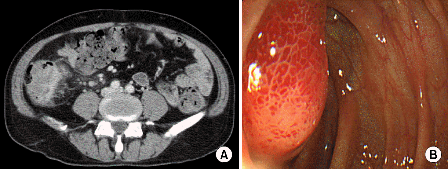 | Figure 2.Abdominal computed tomography scan showed mass- like lesion with contrast enhancement around terminal ileum (A). Followup colonoscopy showed inflammatory polyp at the ileo-cecal valve (B). |
 XML Download