

 PDF
PDF ePub
ePub Citation
 Print
Print


Abstract
Primary Sjögren's syndrome (pSS) is characterized by chronic inflammation and dysfunction in exocrine organs; however, it also has protean clinical features, including neuropsychiatric symptoms. A major neurological manifestation is peripheral neuropathy and involvement of the central nervous system is uncommonly described in pSS. A 52-year-old female was admitted because of depression, dysarthria, gait abnormality, and memory disturbance, which had developed over two months, and was diagnosed as pSS. She was treated successfully with high-dose glucocorticoid and cyclophosphamide pulse therapy without re-currence during the followup period of two years. Herein, we describe the first Korean case of pSS presenting with rapidly progressive cognitive impairment along with a review of the literature.
References
1. Chai J, Logigian EL. Neurological manifestations of primary Sjögren's syndrome. Curr Opin Neurol. 2010; 23:509–13.
2. Segal B, Carpenter A, Walk D. Involvement of nervous system pathways in primary Sjögren's syndrome. Rheum Dis Clin North Am. 2008; 34:885–906.

3. Caselli RJ, Scheithauer BW, Bowles CA, Trenerry MR, Meyer FB, Smigielski JS, et al. The treatable dementia of Sjögren's syndrome. Ann Neurol. 1991; 30:98–101.
4. Kawashima N, Shindo R, Kohno M. Primary Sjögren's syndrome with subcortical dementia. Intern Med. 1993; 32:561–4.
5. Johnstone B, Pepmuelle PH, Vieth AZ, Komatireddy G. Effective treatment of neuropsychological deficits in Sjögren's syndrome. Appl Neuropsychol. 1996; 3:122–7.
6. Michel L, Toulgoat F, Desal H, Laplaud DA, Magot A, Hamidou M, et al. Atypical neurologic complications in patients with primary Sjögren's syndrome: report of 4 cases. Semin Arthritis Rheum. 2011; 40:338–42.
7. Vitali C, Bombardieri S, Jonsson R, Moutsopoulos HM, Alexander EL, Carsons SE, et al. European Study Group on Classification Criteria for Sjögren's Syndrome. Classification criteria for Sjögren's syndrome: a revised version of the European criteria proposed by the American-European Consensus Group. Ann Rheum Dis. 2002; 61:554–8.
8. Shiboski SC, Shiboski CH, Criswell L, Baer A, Challacombe S, Lanfranchi H, et al. Sjögren's International Collaborative Clinical Alliance (SICCA) Research Groups. American College of Rheumatology classification criteria for Sjögren's syndrome: a data-driven, expert consensus approach in the Sjögren's International Collaborative Clinical Alliance cohort. Arthritis Care Res (Hoboken). 2012; 64:475–87.
9. Seo SH, Kim HS, Kwok SK, Ju JH, Kim SH, Yoon CH, et al. Extraglandular manifestations and autoantibodies of Korean patients with primary Sjögren's syndrome. J Korean Rheum Assoc. 2007; 14:43–50.
10. Cho JH, Kim SM, Kim JH, Chu CK, Lee MH, Shin HW, et al. Two cases of primary Sjögren's syndrome presenting as relapsing-remitting multiple sclerosis. J Korean Neurol Assoc. 2004; 22:410–13.
11. Choi HJ, Shin K, Kang EH, Im CH, Lee YJ, Lee EB, et al. Primary Sjögren's syndrome presenting as acute transverse myelitis. Korean J Med. 2005; 68:463–6.
12. Min JH, Kim HJ, Kim BJ, Lee KW, Sunwoo IN, Kim SM, et al. Brain abnormalities in Sjogren syndrome with recurrent CNS manifestations: association with neuromyelitis optica. Mult Scler. 2009; 15:1069–76.
13. Kim MJ, Lee MC, Lee JH, Chung SJ. Cerebellar degener-ation associated with Sjögren's syndrome. J Clin Neurol. 2012; 8:155–9.
14. Delalande S, de Seze J, Fauchais AL, Hachulla E, Stojkovic T, Ferriby D, et al. Neurologic manifestations in primary Sjögren syndrome: a study of 82 patients. Medicine (Baltimore). 2004; 83:280–91.
15. Alexander EL, Ranzenbach MR, Kumar AJ, Kozachuk WE, Rosenbaum AE, Patronas N, et al. Anti-Ro(SS-A) autoantibodies in central nervous system disease associated with Sjögren's syndrome (CNS-SS): clinical, neu-roimaging, and angiographic correlates. Neurology. 1994; 44:899–908.
Figure 1.
Brain MRI demonstrated increased signal intensity on axial T2-weighted images in both cerebral white matter, internal capsules, and cerebellar white matter. These lesions have low signal intensity on axial T1-weighted images and ADC (apparent diffusion coefficient) maps.

Figure 2.
Brain SPECT (single positron emission computed tomography), taken 2 weeks before her admission, reveals resting hypoperfusion of the left frontal, temporal, and parietal cortex and left basal ganglia (arrows).

Figure 3.
Salivary gland scan reveals decreased uptake in the right submandibular gland and excretory dysfunction in both submandibular glands.

Figure 4.
Biopsy of a minor salivary gland showing focal lymphocytic infiltration with a focus score 1 (original magnification, ×400).
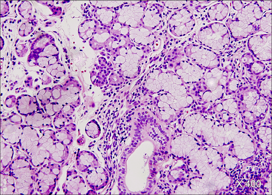
Table 1.
Cases of Sjögren's syndrome presenting cognitive dysfunction responsive to immunosuppresive therapy
| Case 1 (3) | Case 2 (4) | Case 3 (5) | Case 4 (6) | The present case | |
|---|---|---|---|---|---|
| Age (years old) | 56 | 48 | 29 | 57 | 52 |
| Gender | Female | Male | Female | Female | Female |
| Classification criteria of pSS described∗ | Dry mouth (+), Dry eye (+) Schirmer test (+), Anti-Ro/La (+/+) | Dry mouth (+), Dry eye (+) Schirmer test (+), Anti-Ro/La (+/-), MSG† biopsy (+) | Dry mouth (+), Dry eye (+) Schirmer test (+), Anti-Ro/La (+/+), MSG biopsy (+) | Dry mouth (+), Dry eye (+) MSG biopsy (+) | Schirmer test (+), Salivary scan (+), Anti-Ro/La (+/+), MSG biopsy (+) |
| Time from exocrinopathy to CSN symptoms | 15 years | ND‡ | 17 months | ND | - |
| Presenting neurological symptoms | Forgetfulness, Visual hallucination | Lassitude, Forgetfulness, Sterotypic speech | Dissociation, Memory disturbance | y Ataxia, Hypoesthesia, Memory difficulty, Depression | Depression, Dysphagia, Gait disturbance, Memory disturbance |
| MRI | Normal | T2 hyperintense small lesions in the periventricular white matter | Normal | T2 hyperintense lesions in the spinal cord and left frontal white matter | T2 hyperintense lesion, Both cerebellum, cerebrum, internal capsule |
| Treatment | High dose of PD§ (120 mg/day) | High dose of PD (40 mg/day) | High-dose of PD (40 mg/day) | Methylprednisolone pulse and then high dose of PD (1 mg/kg) | Methylprednisone 1 g/day IV and then high dose of PDL¶ (1 mg/kg), CTX∗∗ pulse |
| Follow up | Improvement after 1 month | An episode of relapse after 11 months during PD tapering | Two episodes of relapse during 6 months of Pd tapering | ND | Improvement after 6 weeks No relapse during 2 years |
 XML Download