

 PDF
PDF ePub
ePub Citation
 Print
Print


Abstract
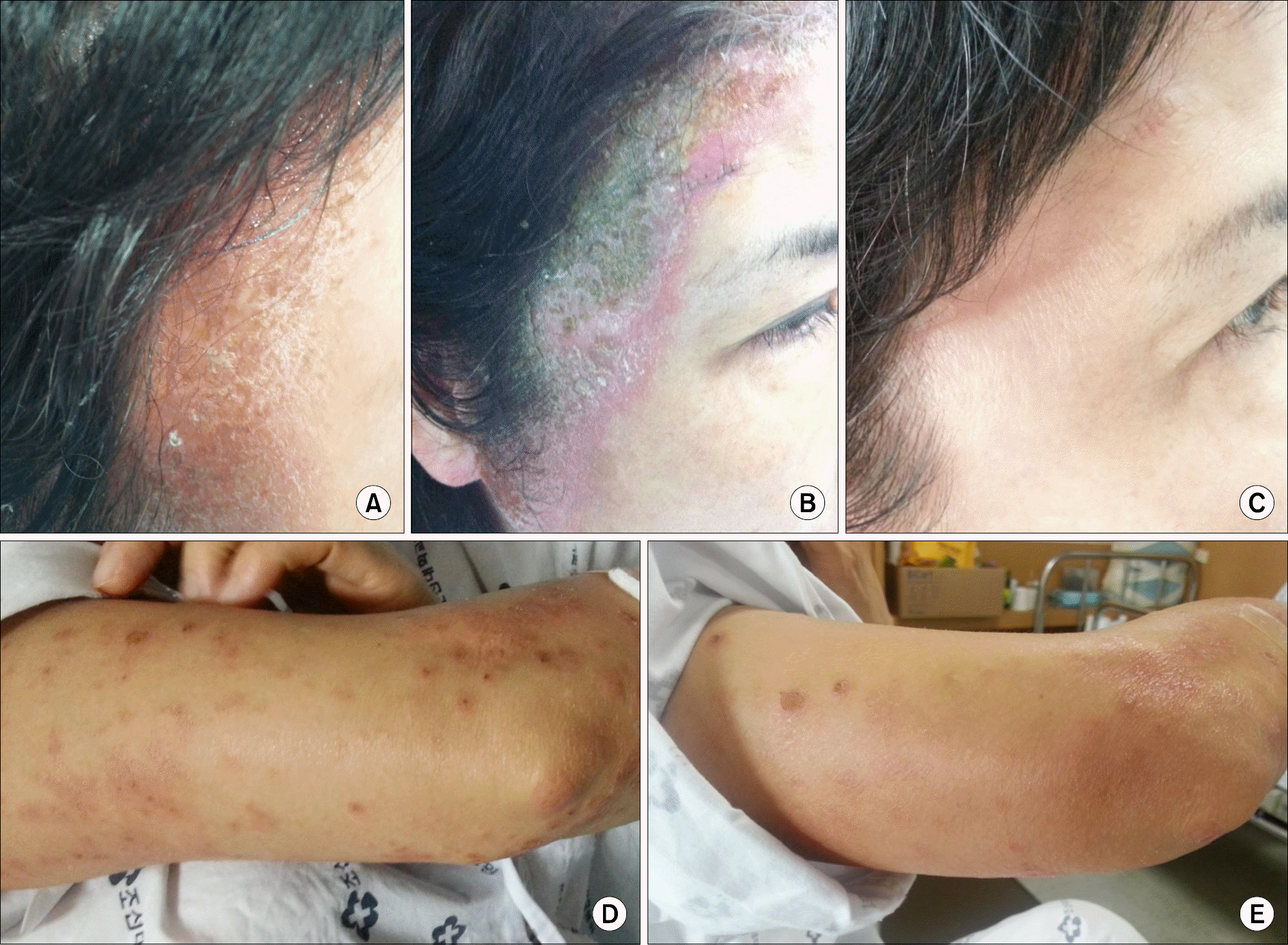
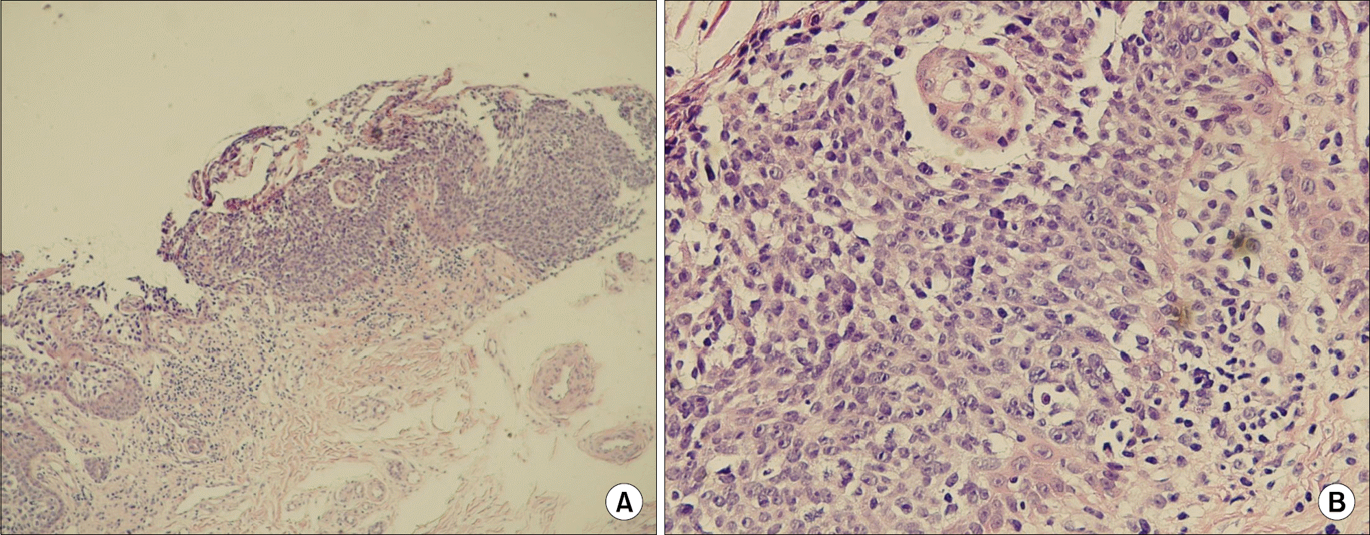
Although tumor necrosis factor (TNF)-α antagonist is a successful treatment modality for various autoimmune diseases, including rheumatoid arthritis (RA), ankylosing spondylitis and psoriatic arthritis, many adverse effects have been reported. Cutaneous adverse reactions of TNF-α antagonist include skin rash, urticaria, lupus like rash, sebor-rheic dermatitis and different kinds of psoriasiform dermatitis. We report a case of psoriasiform dermatitis during TNF-α antagonist treatment in a 50-year-old woman with RA. The patient has been treated with adalimumab. After 2 months, she developed pruritic erythematous eruption and desquamative lesions on the head and limbs, which were defined as psoriasiform change by a skin biopsy. These skin lesions are successfully treated with combination therapy, including cessation of adalimumab, corticosteroid and phototherapy.
References
1. Bongartz T, Sutton AJ, Sweeting MJ, Buchan I, Matteson EL, Montori V. Anti-TNF antibody therapy in rheumatoid arthritis and the risk of serious infections and malignancies: systematic review and meta-analysis of rare harmful effects in randomized controlled trials. JAMA. 2006; 295:2275–85.
2. Moustou AE, Matekovits A, Dessinioti C, Antoniou C, Sfikakis PP, Stratigos AJ. Cutaneous side effects of antitumor necrosis factor biologic therapy: a clinical review. J Am Acad Dermatol. 2009; 61:486–504.

3. Choi YJ, Kim DS, Park JM, Oh SH, Park YK, Lee JH. A case of psoriasiform eruption triggered by tumor necrosis factor-alpha antagonist therapy. Korean J Dermatol. 2008; 46:721–3.
4. Oh JM, Koh EM, Kim H, Lee J, Ahn JK, Cha HS, et al. Exacerbation of psoriatic skin lesion followed by tnf-alpha antagonist treatment. J Korean Rheum Assoc. 2010; 17:200–4.
5. Mease P. TNFalpha therapy in psoriatic arthritis and psoriasis. Ann Rheum Dis. 2004; 63:755–8.
6. Alsaad KO, Ghazarian D. My approach to superficial inflammatory dermatoses. J Clin Pathol. 2005; 58:1233–41.
7. Mehta S, Singal A, Singh N, Bhattacharya SN. A study of clinicohistopathological correlation in patients of psoriasis and psoriasiform dermatitis. Indian J Dermatol Venereol Leprol. 2009; 75:100–10.
8. Schopf RE, Aust H, Knop J. Treatment of psoriasis with the chimeric monoclonal antibody against tumor necrosis factor alpha, infliximab. J Am Acad Dermatol. 2002; 46:886–91.
9. Fouache D, Goëb V, Massy-Guillemant N, Avenel G, Bacquet-Deschryver H, Kozyreff-Meurice M, et al. Paradoxical adverse events of anti-tumour necrosis factor therapy for spondyloarthropathies: a retrospective study. Rheumatology (Oxford). 2009; 48:761–4.
10. Sfikakis PP, Iliopoulos A, Elezoglou A, Kittas C, Stratigos A. Psoriasis induced by antitumor necrosis factor therapy: a paradoxical adverse reaction. Arthritis Rheum. 2005; 52:2513–8.
11. de Gannes GC, Ghoreishi M, Pope J, Russell A, Bell D, Adams S, et al. Psoriasis and pustular dermatitis triggered by TNF-{alpha} inhibitors in patients with rheumatologic conditions. Arch Dermatol. 2007; 143:223–31.
12. Collamer AN, Guerrero KT, Henning JS, Battafarano DF. Psoriatic skin lesions induced by tumor necrosis factor antagonist therapy: a literature review and potential mechanisms of action. Arthritis Rheum. 2008; 59:996–1001.
 XML Download