

 PDF
PDF ePub
ePub Citation
 Print
Print


Abstract
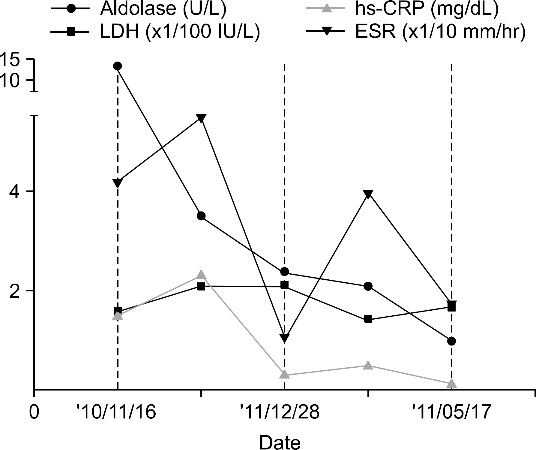
We describe a case of localized gastrocnemius myositis which developed with flare-up of Crohn's disease. A 21-year old male patient with an 8-year history of Crohn's disease presented with pain and tenderness in both calves without recent abdominal symptoms. Electromyography and gastrocnemius muscle biopsy revealed evidence of inflammatory myositis. Magnetic resonance imaging (MRI) showed bilateral symmetrical diffuse increased signal intensity in T2 weighted images in both gastrocnemius muscles and patchy contrast enhancement. Subsequent gastrointestinal investigation revealed active inflammation of colon with multiple pseudopolyps and enteroenteric fistula on which we com-menced oral prednisolone of 30 mg daily. His pain on both calves was improved and muscle enzymes became normal. Following dose reduction of prednisolone, azathioprine 50 mg daily was started considering the patient's active Crohn's disease on endoscopic findings prior to the development of overt abdominal symptoms. This is the first case report of localized gastrocnemius myositis associated with Crohn's disease described in Korea. Calf myositis re-sponded to corticosteroid well and did not recur with maintenance therapy using azathioprine and mesalazine.
References
1. Bourikas LA, Papadakis KA. Musculoskeletal manifestations of inflammatory bowel disease. Inflamm Bowel Dis. 2009; 15:1915–24.

2. Mogul Z, Katz S, Bachman TR, Urmacher C. Isolated gastrocnemius myositis related to Crohn's disease. Gastroenterol Hepatol (NY). 2010; 6:453–5.
3. Christopoulos C, Savva S, Pylarinou S, Diakakis A, Papavassiliou E, Economopoulos P. Localised gastrocnemius myositis in Crohn's disease. Clin Rheumatol. 2003; 22:143–5.
4. Szabo N, Lukacs S, Kulcsar I, Gunasekera W, Nagy-Toldi A, Dezso B, et al. Association of idiopathic inflammatory myopathy and Crohn's disease. Clin Rheumatol. 2009; 28:99–101.
5. Shimoyama T, Tamura Y, Sakamoto T, Inoue K. Immune-mediated myositis in Crohn's disease. Muscle Nerve. 2009; 39:101–5.
6. Orlando A, Modesto I, Castiglione F, Scala L, Scimeca D, Rispo A, et al. The role of calprotectin in predicting endoscopic post-surgical recurrence in asymptomatic Crohn's disease: a comparison with ultrasound. Eur Rev Med Pharmacol Sci. 2006; 10:17–22.
7. Manganelli S, De Stefano R, Malandrini A, Selvi E, Frati E, Gambelli S, et al. Bilateral recurrent focal myositis of gastrocnemius muscles after BCG vaccination. Rheumatology (Oxford). 2002; 41:1074–6.
Figure 1.
Both lower leg MRI showed bilateral symmetrical diffuse signal alteration of gastrocnemius muscles and patchy contrast enhancement (arrow) consis-tent with inflammatory myositis.
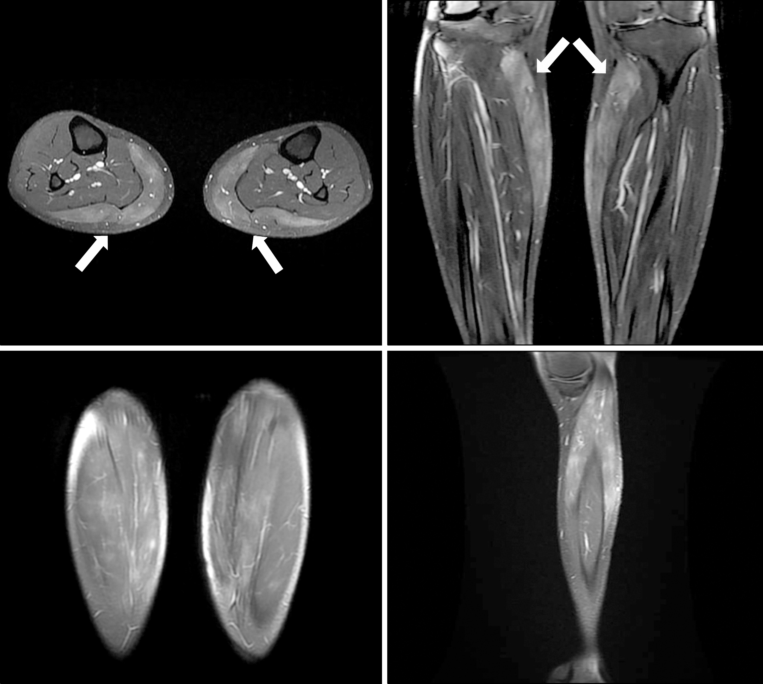
Figure 2.
Biopsy of the left lateral gastrocnemius muscle revealed that there is a mild size variation of myofibers and inflammatory cell infiltration in the endomysium and perivsacular area. Some degenerating and regenerating myofibers are shown. Internal nuclei are not prominent. There is mild fibrosis and fat ingrowth in the endomysium but no evidence of vasculitis.
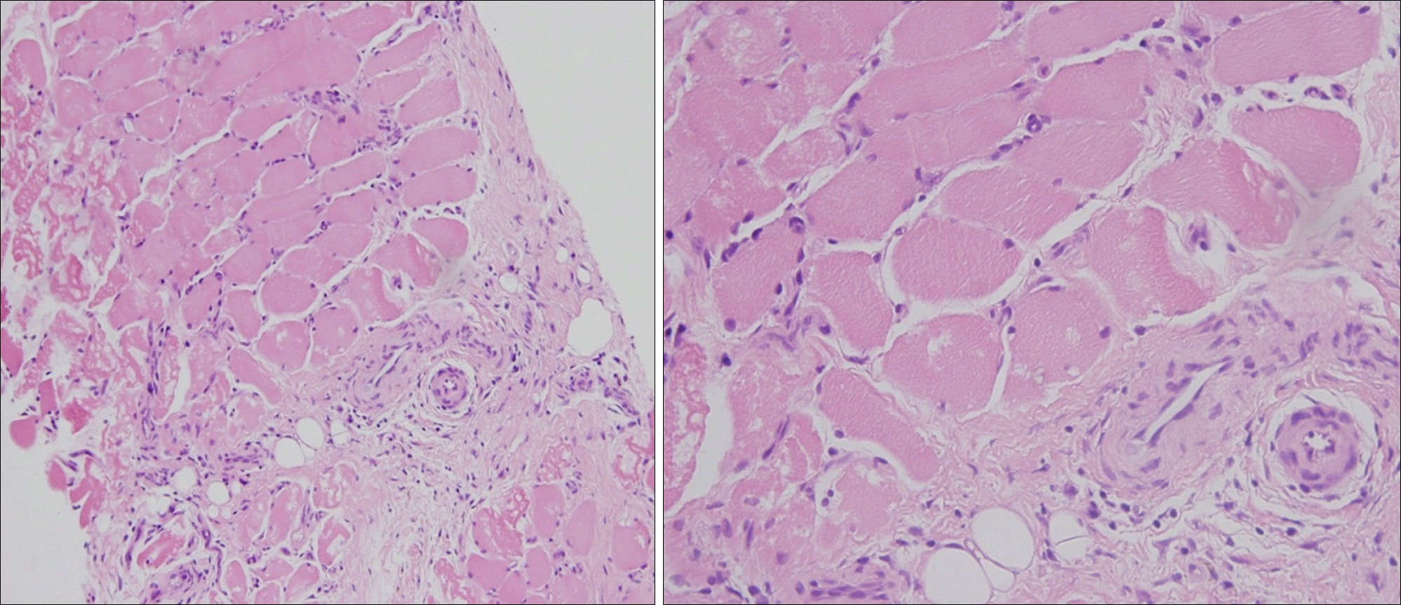
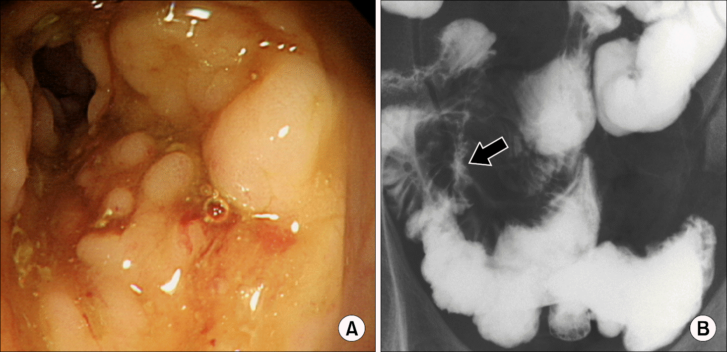
Figure 3.
(A) Colonoscopy showing cobble stone appearance over cecum, IC valve, terminal ileum and longitudinal ulcer along the mesenteric border of terminal ileum were observed. (B) Small bowel barium enema showing multifocal pseudosacculation in the antime-senteric border with interpositio-ning of the normal ileum was found. Irregular contrast leakage in the right lower quadrant area were visible, apparently suggesting enteroenteric fistula confined to me-sentery (arrow).
 XML Download