

 PDF
PDF ePub
ePub Citation
 Print
Print


Abstract
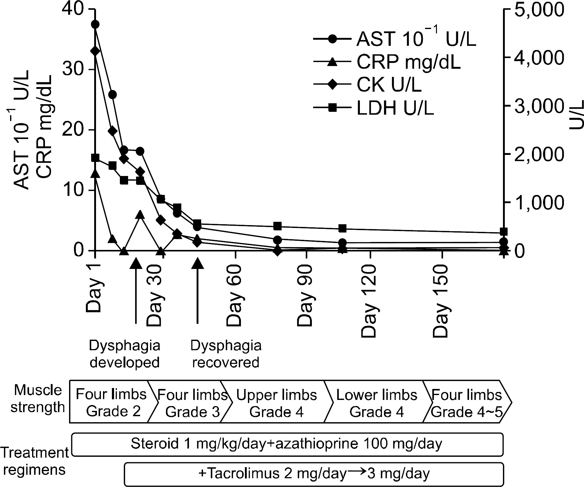
An overlap syndrome is a combination of major features of more than one connective tissue diseases which is presented in the same patient. An overlap syndrome of rheumatoid arthritis (RA) and polymyositis (PM) which involved the upper pharyngeal muscle has not been reported in Korea. Herein, we report a rare case of a patient with a long-history RA presenting proximal muscle weakness and swallowing difficulty, who was successfully treated with a high-dose of corticosteroid, azathioprine and tacrolimus.
References
1. Cervera R, Khamashta MA, Hughes GR. ‘Overlap' syndromes. Ann Rheum Dis. 1990; 49:947–8.

2. Soriano ER, McHugh NJ. Overlap syndromes in adults and children. Madison PJ, Isenberg DA, Woo P, Glass DN, editors. Oxford rheumatology. 2nd ed.p. 1413–32. New York: Oxford University press;1998.
3. Martínez-Cordero E, León DE, Ortega LA. Association of polymyositis with rheumatoid arthritis. Rheumatol Int. 2001; 20:119–23.
4. Agrawal V, Husain N, Das SK, Bagchi M. Muscle involvement in rheumatoid arthritis: clinical and histological characteristic and review of literature. J Indian Rheumatol Assoc. 2003; 11:98–103.
5. Hur JW, Lee CW, Yoo DH. Bucillamine-induced pemphi-gus vulgaris in a patient with rheumatoid arthritis and polymyositis overlap syndrome. J Korean Med Sci. 2006; 21:585–7.
6. Lim HW, Shin JN, Kang HW, Yoo JW, Kim J. A case of overlap syndrome with rheumatoid arthritis and polymyositis. J Korean Rheum Assoc. 2006; 13:64–9.
7. Sharp GC, Irvin WS, Tan EM, Gould RG, Holman HR. Mixed connective tissue disease–an apparently distinct rheumatic disease syndrome associated with a specific antibody to an extractable nuclear antigen (ENA). Am J Med. 1972; 52:148–59.
8. Marie I, Mouthon L. Therapy of polymyositis and dermatomyositis. Autoimmun Rev. 2011; 11:6–13.
9. Marie I, Menard JF, Hatron PY, Hachulla E, Mouthon L, Tiev K, et al. Intravenous immunoglobulins for steroid-refractory esophageal involvement related to polymyositis and dermatomyositis: a series of 73 patients. Arthritis Care Res (Hoboken). 2010; 62:1748–55.
10. Oddis CV, Sciurba FC, Elmagd KA, Starzl TE. Tacrolimus in refractory polymyositis with interstitial lung disease. Lancet. 1999; 353:1762–3.
11. Schumacher HR, Schimmer B, Gordon GV, Bookspan MA, Brogadir S, Dorwart BB. Articular manifestations of polymyositis and dermatomyositis. Am J Med. 1979; 67:287–92.
12. Steiner G. Autoantibody in rheumatoid arthritis. Hochberg MC, Silman AJ, Smolen JS, editors. Rheumatology. 3rd ed.p. 833–41. London: Mosby;2003.
13. Stone KB, Oddis CV, Fertig N, Katsumata Y, Lucas M, Vogt M, et al. Anti-Jo-1 antibody levels correlate with disease activity in idiopathic inflammatory myopathy. Arthritis Rheum. 2007; 56:3125–31.
14. Sanmartí R, Collado A, Gratacós J, Bedini JL, Pañella D, Filella X, et al. Reduced activity of serum creatine kinase in rheumatoid arthritis: a phenomenon linked to the inflammatory response. Br J Rheumatol. 1994; 33:231–4.
15. Nagashima T, Iwamoto M, Minota S. Moderate incidence of prior rheumatoid arthritis in patients with polymyositis and dermatomyositis. Clin Rheumatol. 2011; 30:875–6.
Figure 1.
Both hand radiographs show characteristic features of rheumatoid arthritis, including periarticular osteopenia, joint space narrowing and articular erosions of carpometacarpal, radiocarpal and intercarpal joints.
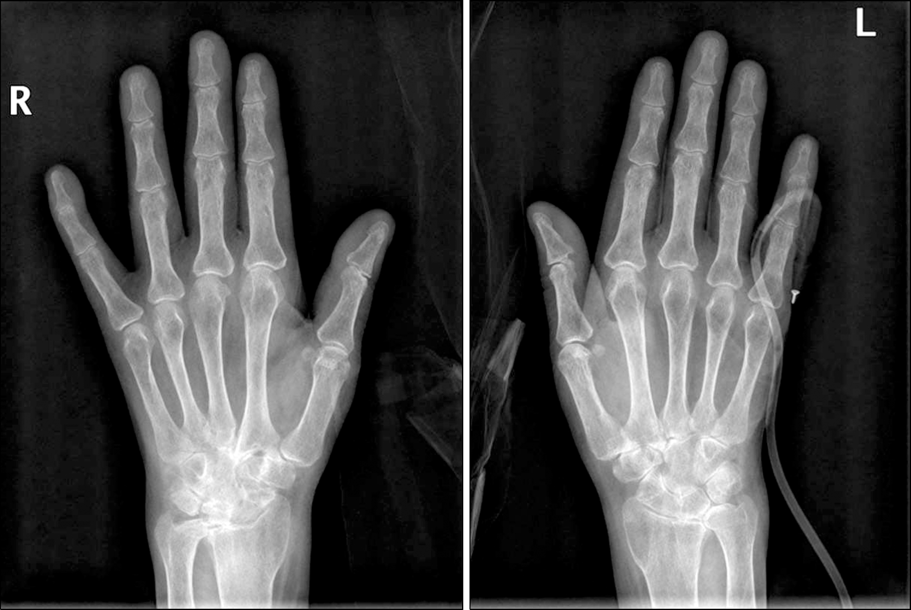
Figure 2.
Radiographic changes typical of rheumatoid arthritis on anteroposterior foot X-ray. This revealed bony erosions with joint space narrowing of both metatarsophalangeal, intertarsal and tarsometatarsal joints.
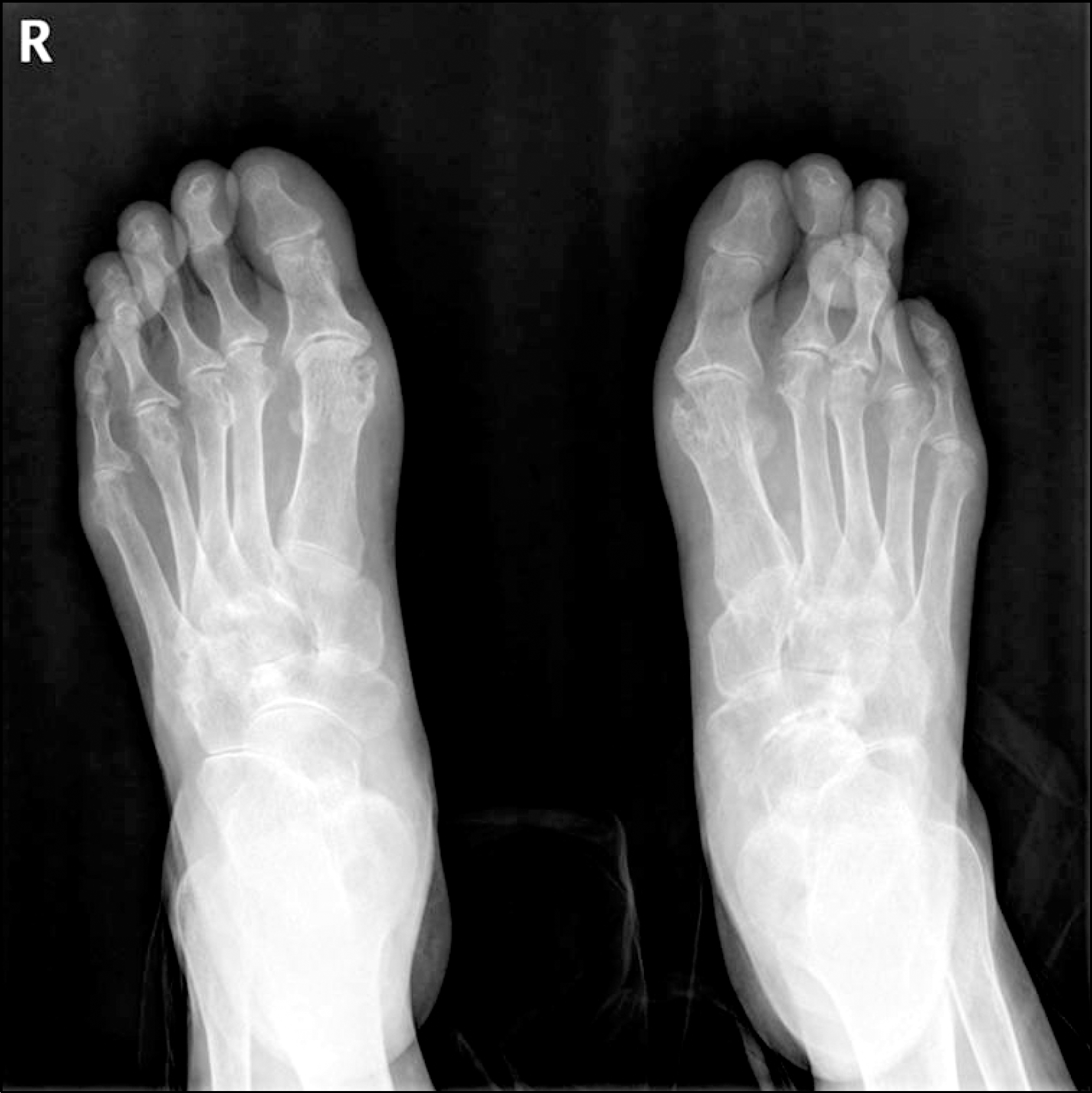
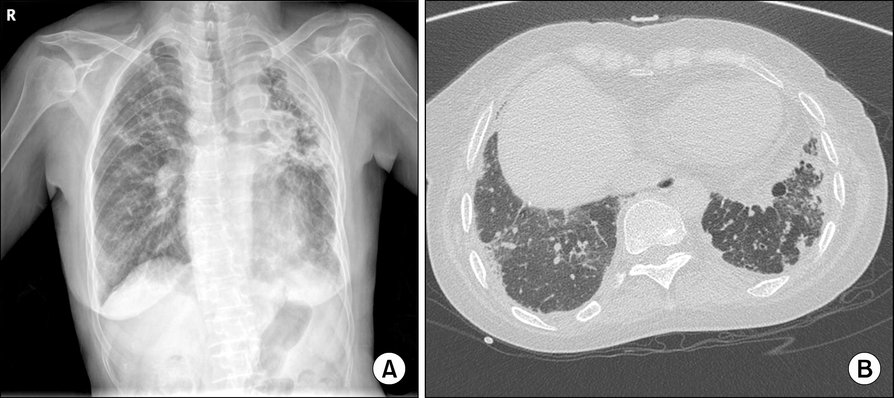
Figure 3.
Initial chest radiograph shows characteristic findings of a “tuberculosis-destroyed lung” (A). A chest computed tomography revealed interstitial lung disease at the basal portion of the right lower lobe (B).
 XML Download