

 PDF
PDF ePub
ePub Citation
 Print
Print


Abstract
Primary hypertrophic osteoarthropathy or pachydermoperiostosis is a rare hereditary disorder characterized by digital clubbing, pachydermia and periostosis. Its precise incidence and prevalence is still unknown due to the lack of controlled data. It occurs without any underlying causes and usually has a chronic course. Life expectancy may be of normal standards, but many patients develop multiple functional and cosmetic complications. So, it is important to diagnose this disease at an early stage and to treat the symptomat for the quality of life. We report a case of primary hypertrophic osteoarthropathy in a 68-year-old male with clinical features such as digital clubbing and pachydermia, radiographic findings of acroosteolysis and periosteal new bone formation.
Go to : 
References
1. Friedreich N. Hyprstose es gesmmten skelettes. Arh Pathol Ant. 1868; 43:83–7.
2. Castori M, Sinibaldi L, Mingarelli R, Lachman RS, Rimoin DL, Dallapiccola B. Pachydermoperiostosis: an update. Clin Genet. 2005; 68:477–86.

3. Martí nez-Lavín M, Vargas AS, Cabré J, Nava A, Silveira LH, Amigo MC, et al. Features of hypertrophic osteoarthropathy in patients with POEMS syndrome: a metaanalysis. J Rheumatol. 1997; 24:2267–8.
4. Jajic Z, Jajic I, Nemcic T. Primary hypertrophic osteoarthropathy: clinical, radiologic, and scintigraphic characteristics. Arch Med Res. 2001; 32:136–42.
5. Touraine A, Solente G, Golé L. Un syndrome osteo-dermopathique: la pachydermie plicaturee avec pachyper-ostose des extremites. Presse Med. 1935; 43:1820–4.
6. Silveira LH, Martí nez-Lavín M, Pineda C, Fonseca MC, Navarro C, Nava A. Vascular endothelial growth factor and hypertrophic osteoarthropathy. Clin Exp Rheumatol. 2000; 18:57–62.
7. Poormoghim H, Hosseynian A, Javadi A. Primary hypertrophic osteoarthropathy. Rheumatol Int. 2012; 32:607–10.
8. Paik MH, Lee BY, Lee KH, Song KS, Kim JW, Min KO. Pachydermoperiostosis (primary hypertrophic osteoarthropathy): case report. J Korean Radiol Soc. 2002; 47:533–8.
9. Pineda CJ, Guerra J Jr, Weisman MH, Resnick D, Martinez-Lavin M. The skeletal manifestations of clubbing: a study in patients with cyanotic congenital heart disease and hypertrophic osteoarthropathy. Semin Arthritis Rheum. 1985; 14:263–73.
10. Hedayati H, Barmada R, Skosey JL. Acrolysis in pachydermoperiostosis. Primary or idiopathic hypertrophic osteoarthropathy. Arch Intern Med. 1980; 140:1087–8.
11. Joseph B, Chacko V. Acroosteolysis associated with hypertrophic pulmonary osteoarthropathy and pachydermoperiostosis. Radiology. 1985; 154:343–4.
12. Lee J, Kim H, Hwang JW, Noh JW, Ahn JK, Koh EM, et al. Arthroscopic synovectomy in a patient with primary hypertrophic osteoarthropathy. J Korean Rheum Assoc. 2008; 15:261–7.
13. Cooper RG, Freemont AJ, Riley M, Holt PJ, Anderson DC, Jayson MI. Bone abnormalities and severe arthritis in pachydermoperiostosis. Ann Rheum Dis. 1992; 51:416–9.
14. Kwon MH, Joung CI. Pachydermoperiostosis mimicking ac-romegaly: A case report. Turk J Rheumatol. 2012; 27:132–5.
15. Hong YS, Yang HI, Park SH, Lee SH, Cho CS, Kim HY. A case of Sjogren syndrome associated with acroosteolysis. J Korean Rheum Assoc. 1996; 3:92–6.
Go to :
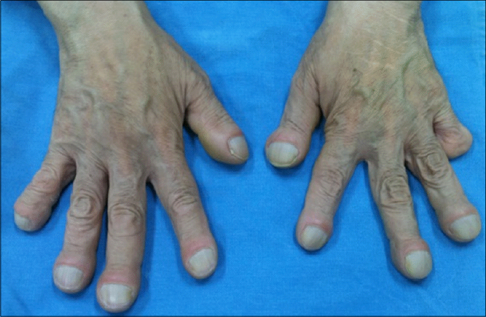 | Figure 1.Digital clubbing on both hands and the amputated 5 th mid phalanx of left hand are seen. |
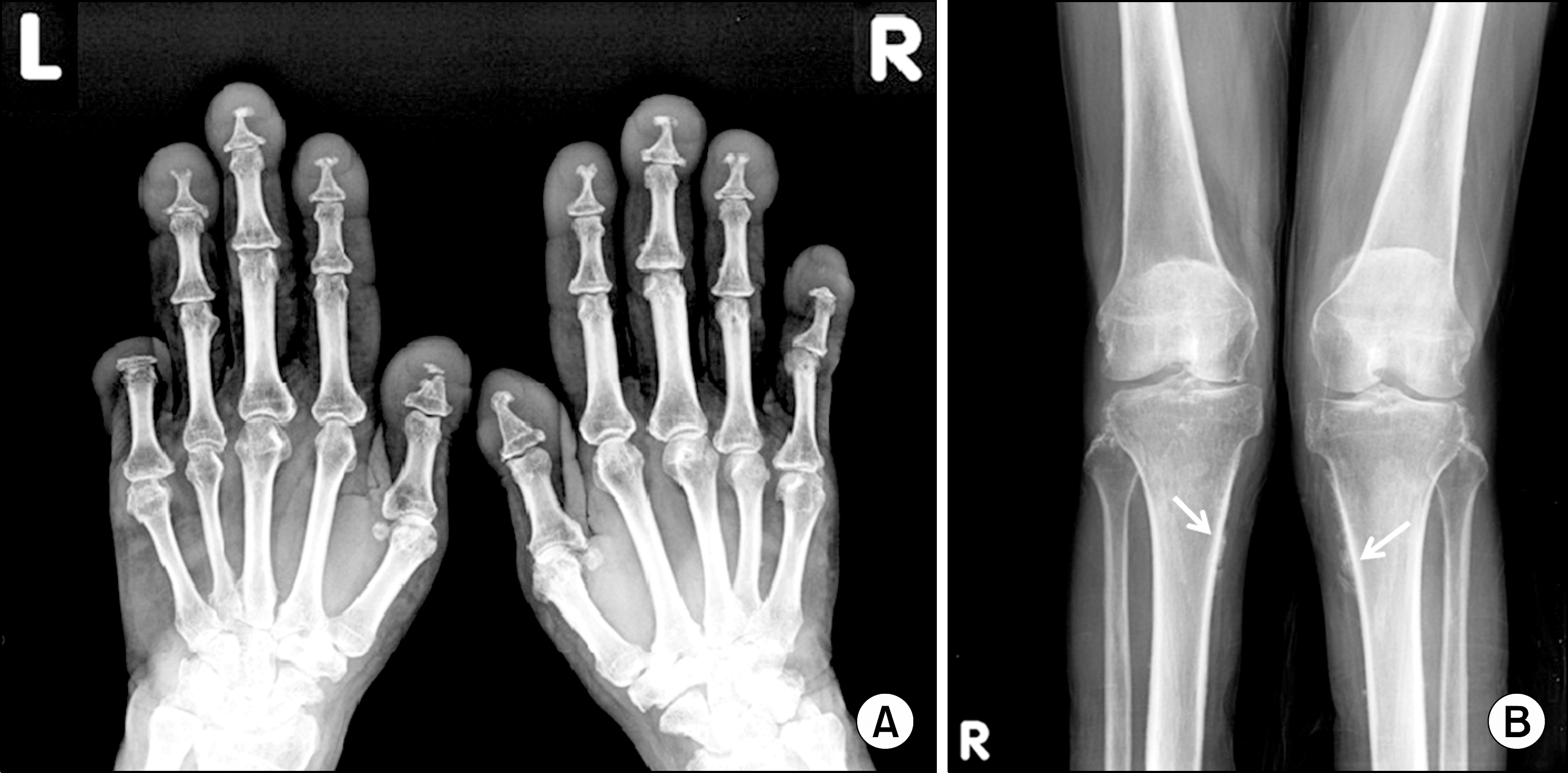 | Figure 2.(A) Plain radiograph of both hands demonstrate acroosteolysis. Extensive bone lysis of distal phalanges with bases for the preservation are seen. (B) Plain radiograph of lower extremities shows irregular linear cortical thickening of both proximal tibia (white arrows) and periosteal new bone formation. |
 XML Download