Abstract
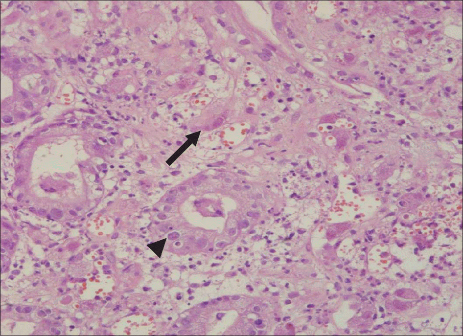
Cytomegalovirus (CMV) is a relatively common viral patho-gen, and CMV infection is generally assumed asymptomatic in general hosts. In immunologically compromised patients, CMV infection can cause further serious diseases such as pneumonitis, retinitis, encephalitis, and enterocolitis. A 40-year-old man is being presented with acute fever, myal-gia, and sore throat. Laboratory findings have revealed elevated ESR, CRP, and ferritin levels. The patient was being treated for adult-onset Still's disease (AOSD). Three weeks later, although AOSD activity was under control, the patient began to complain about oral soreness, epigastric pain, and diarrhea. Endoscopy revealed multiple round ulcers with white patches in the esophagus and the stomach, sparing the colon. Anti-fungal agent is being administered but failed to bring improvements after 2 weeks of therapy. CMV infection is confirmed with pathology, antiviral agents were initiated after the ulcers subsided. Currently, clinical associations between CMV infection and AOSD are suggested. CMV infection may be considered as a differential diagnosis when multiple upper gastrointestinal ulcerative lesions develop within patients whom have been treated AOSD with immunosuppressive agents.
References
1. Himoto T, Goda F, Okuyama H, Kono T, Yamagami A, Inukai M, et al. Cytomegalovirus-associated acute gastric mucosal lesion in an immunocompetent host. Intern Med. 2009; 48:1521–4.

2. Ishiguro T, Takayanagi N, Kawabata Y, Sugita Y. Intestinal perforation due to concomitant cytomegalovirus infection during treatment for Pneumocystis jirovecii pneumonia in a patient with rheumatoid arthritis. Intern Med. 2011; 50:1835–7.

3. Canbakan B, Mizrak D, Keven K, Kaygusuz G, Kutlay S, Sengul S, et al. Gastric ulcer despite no acid in a renal allograft recipient: what is the link? Nephrol Dial Transplant. 2005; 20:2279–81.

4. Ju JH, Park HS, Shin MJ, Yang CW, Kim YS, Choi YJ, et al. Successful treatment of massive lower gastrointestinal bleeding caused by mixed infection of cytomegalovirus and mucormycosis in a renal transplant recipient. Am J Nephrol. 2001; 21:232–6.

5. Péter A, Telkes G, Varga M, Sá rváry E, Kovalszky I. Endoscopic diagnosis of cytomegalovirus infection of upper gastrointestinal tract in solid organ transplant recipi-ents: Hungarian single-center experience. Clin Transplant. 2004; 18:580–4.

6. Wilcox CM, Chalasani N, Lazenby A, Schwartz DA. Cytomegalovirus colitis in acquired immunodeficiency syndrome: a clinical and endoscopic study. Gastrointest Endosc. 1998; 48:39–43.

7. Hirsch MS. Cytomegalovirus and Human Herpesvirus Types 6, 7, and 8. 18th ed.p. 1471–5. McGraw Hill;2012.
8. Lin WR, Su MY, Hsu CM, Ho YP, Ngan KW, Chiu CT, et al. Clinical and endoscopic features for alimentary tract cytomegalovirus disease: report of 20 cases with gastrointestinal cytomegalovirus disease. Chang Gung Med J. 2005; 28:476–84.
9. Patra S, Samal SC, Chacko A, Mathan VI, Mathan MM. Cytomegalovirus infection of the human gastrointestinal tract. J Gastroenterol Hepatol. 1999; 14:973–6.

10. Bento DP, Tavares R, Baptista Leite R, Miranda A, Ramos S, Ventura F, et al. Adult-Onset Still's Disease and cytomegalovirus infection. Acta Reumatol Port. 2010; 35:259–63.
11. Chacko B, John GT. Leflunomide for cytomegalovirus: bench to bedside. Transpl Infect Dis. 2012; 14:111–20.

12. Alanazi AH, Aldekhail WM, Jewell L, Huynh HQ. Multiple large gastric ulcers as a manifestation of cytomegalovirus infection in a healthy child. J Pediatr Gastroenterol Nutr. 2009; 49:364–7.

13. Yoda Y, Hanaoka R, Ide H, Isozaki T, Matsunawa M, Yajima N, et al. Clinical evaluation of patients with inflammatory connective tissue diseases complicated by cytomegalovirus antigenemia. Mod Rheumatol. 2006; 16:137–42.

14. Efthimiou P, Paik PK, Bielory L. Diagnosis and management of adult onset Still's disease. Ann Rheum Dis. 2006; 65:564–72.

15. Amenomori M, Migita K, Miyashita T, Yoshida S, Ito M, Eguchi K, et al. Cytomegalovirus-associated hemophago-cytic syndrome in a patient with adult onset Still's disease. Clin Exp Rheumatol. 2005; 23:100–2.
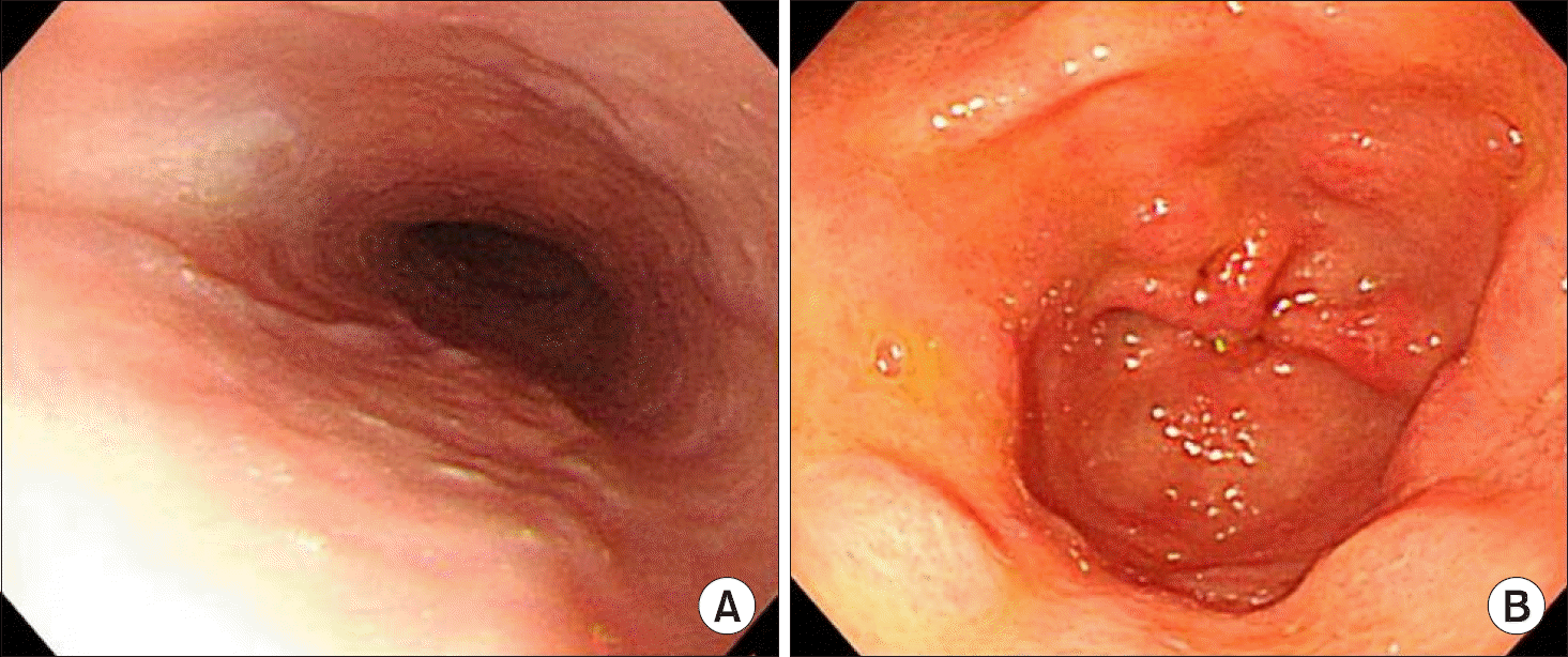
Figure 1.
Gastrointestinal tract endoscopic findings. Multiple whitish plaques are observed in esophagus (A), variable-sized multiple superficial ulcers and erosions are shown in gastric antrum (B), and relatively normal-appearing colonic mucosa is shown (C).


 PDF
PDF ePub
ePub Citation
 Print
Print


 XML Download