

 PDF
PDF ePub
ePub Citation
 Print
Print


Abstract
Polymyalgia rheumatica (PMR) is an inflammatory rheumatic condition characterized by generalized pain and morning stiffness in the shoulders, hip girdle, and neck. Since the pathogenesis of PMR is still uncertain, the diagnosis of PMR depends on clinical features. There have been several studies regarding radiological tools for the diagnosis of PMR. Recent studies using 18-FDG-PET showed bursitis, synovitis, uptake in the spinous process and asymptomatic large-vessel vasculitis in PMR patients. However, there was no report on the efficacy of 18-FDG-PET for diagnosis of PMR in Korea. Here, we are first reporting a case of a Korean patient with PMR, who had radiological findings including bursitis, synovitis, uptake in the spinous process and asymptomatic large-vessel vasculitis on 18-FDG-PET/CT.
Go to : 
References
1. Healey LA. Longterm followup of polymyalgia rheumatica: evidence for synovitis. Semin Arthritis Rheum. 1984; 13:322–8.

2. Salvarani C, Cantini F, Hunder GG. Polymyalgia rheumatica and giant-cell arteritis. Lancet. 2008; 372:234–45.
3. Camellino D, Cimmino MA. Imaging of polymyalgia rheumatica: indications on its pathogenesis, diagnosis and prognosis. Rheumatology (Oxford). 2012; 51:77–86.
4. Blockmans D, De Ceuninck L, Vanderschueren S, Knockaert D, Mortelmans L, Bobbaers H. Repetitive 18-fluorodeoxyglucose positron emission tomography in isolated polymyalgia rheumatica: a prospective study in 35 patients. Rheumatology (Oxford). 2007; 46:672–7.
5. Hoh CK, Schiepers C, Seltzer MA, Gambhir SS, Silverman DH, Czernin J, et al. PET in oncology: will it replace the other modalities? Semin Nucl Med. 1997; 27:94–106.
6. Yamashita H, Kubota K, Takahashi Y, Minaminoto R, Morooka M, Ito K, et al. Whole-body fluorodeoxyglucose positron emission tomography/computed tomography in patients with active polymyalgia rheumatica: evidence for distinctive bursitis and large-vessel vasculitis. Mod Rheumatol. 2012; 22:705–11.
7. Moosig F, Czech N, Mehl C, Henze E, Zeuner RA, Kneba M, et al. Correlation between 18-fluorodeoxyglu-cose accumulation in large vessels and serological markers of inflammation in polymyalgia rheumatica: a quantitative PET study. Ann Rheum Dis. 2004; 63:870–3.
8. Blockmans D, Stroobants S, Maes A, Mortelmans L. Positron emission tomography in giant cell arteritis and polymyalgia rheumatica: evidence for inflammation of the aortic arch. Am J Med. 2000; 108:246–9.
9. Blockmans D, Maes A, Stroobants S, Nuyts J, Bormans G, Knockaert D, et al. New arguments for a vasculitic nature of polymyalgia rheumatica using positron emission tomography. Rheumatology (Oxford). 1999; 38:444–7.
10. Salvarani C, Cantini F, Boiardi L, Hunder GG. Polymyalgia rheumatica. Best Pract Res Clin Rheumatol. 2004; 18:705–22.
11. Sidhom OA, Basalaev M, Sigal LH. Renal cell carcinoma presenting as polymyalgia rheumatica. Resolution after nephrectomy. Arch Intern Med. 1993; 153:2043–5.
12. Brooks RC, McGee SR. Diagnostic dilemmas in polymyalgia rheumatica. Arch Intern Med. 1997; 157:162–8.
Go to :
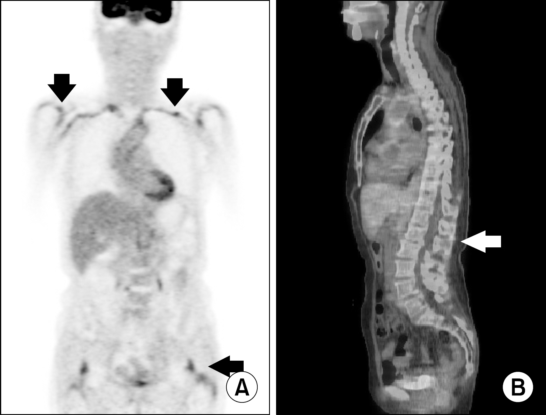 | Figure 2.Multifocal increased FDG uptakes noted at the wall of both subclavian arteries and bursae around both shoulder and hip joints (A). Also focal increased FDG uptakes noted at the interspinous ligaments of lumbar spine (B). |
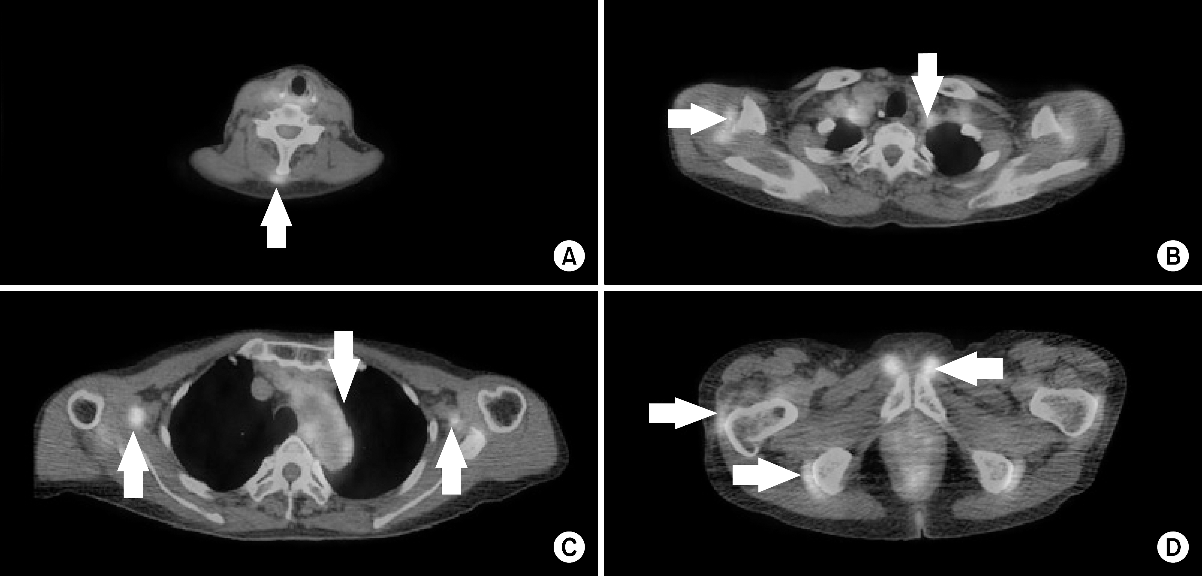 | Figure 3.There are increased FDG uptakes at the interspinous ligaments of cervical spine (A), bursae around both shoulders, both subclavian arteries (B), both brachial arteries and thoracic aorta (C). Also noted increased FDG uptakes at bursae around both greater trochanters, ischial tuberosities and symphysis pubis (D). |
 XML Download