

 PDF
PDF ePub
ePub Citation
 Print
Print


Abstract
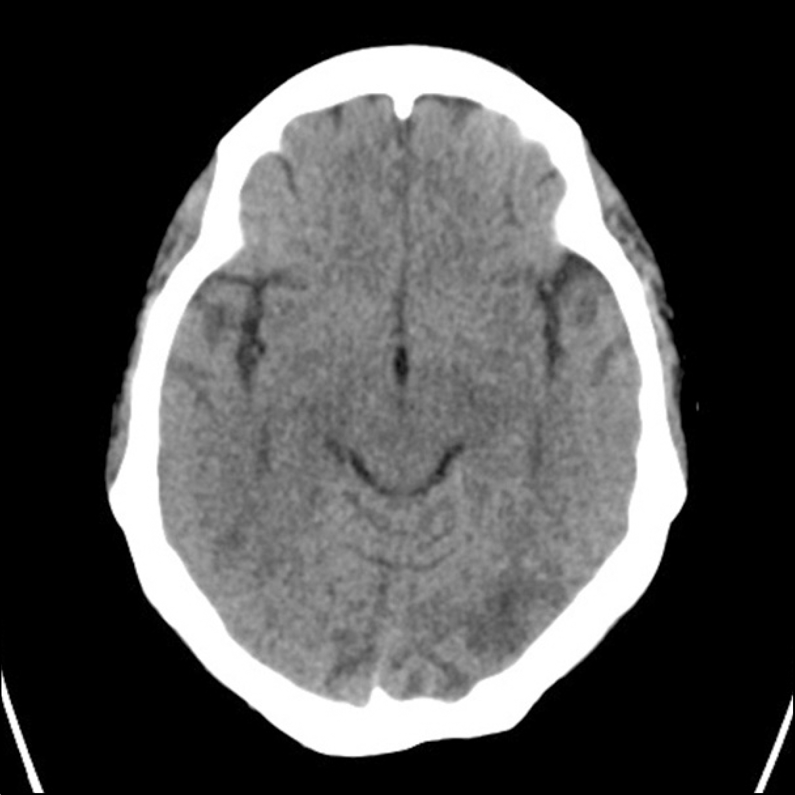
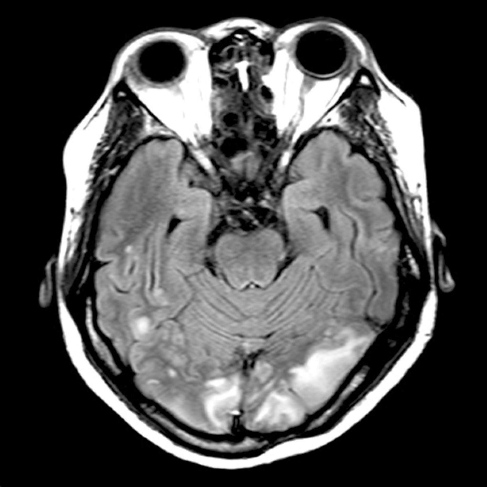
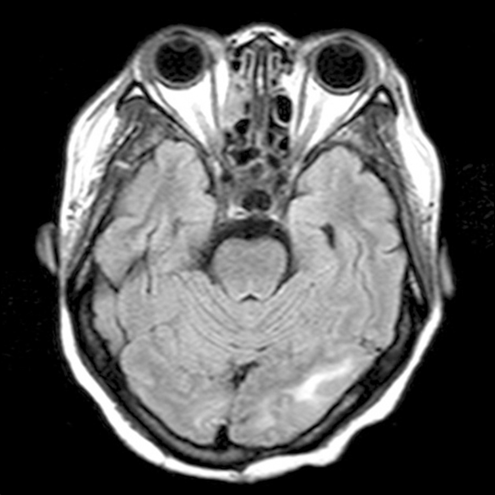
Posterior reversible encephalopathy syndrome (PRES) is a neurologic condition characterized by vasogenic edema on neuroimaging and is associated with the setting of severe hypertension, eclampsia, autoimmune disease, malignancy, and immunosuppressive drugs. We report on a 42 year-old female systemic lupus erythematous patient who presented altered consciousness, seizure, and visual disturbance after cyclophosphamide pulse therapy. Magnetic resonance imaging (MRI) showed multifocal high signal intensity lesions in the parieto-occipital cortex bilaterally and in the subcortical white matter. Her condition was improved and her MRI lesions were resolved after aggressive blood pressure control and high-dose steroid treatment. It is possibly the first reported case of PRES in a patient with lupus, treated with cyclophosphamide pulse therapy during a nephritis flare in Korea.
References
1. Hinchey J, Chaves C, Appignani B, Breen J, Pao L, Wang A, et al. A reversible posterior leukoencephalopathy syndrome. N Engl J Med. 1996; 334:494–500.

2. Casey SO, Sampaio RC, Michel E, Truwit CL. Posterior reversible encephalopathy syndrome: utility of fluid-atte-nuated inversion recovery MR imaging in the detection of cortical and subcortical lesions. AJNR Am J Neuroradiol. 2000; 21:1199–206.
3. Baizabal-Carvallo JF, Barragán-Campos HM, Padilla-Aranda HJ, Alonso-Juarez M, Estañol B, Cantú-Brito C, et al. Posterior reversible encephalopathy syndrome as a complication of acute lupus activity. Clin Neurol Neurosurg. 2009; 111:359–63.
4. Varaprasad IR, Agrawal S, Prabu VN, Rajasekhar L, Kanikannan MA, Narsimulu G. Posterior reversible encephalopathy syndrome in systemic lupus erythematosus. J Rheumatol. 2011; 38:1607–11.
5. Cuadrado MJ, Sanna G. Headache and systemic lupus erythematosus. Lupus. 2003; 12:943–6.
6. Leroux G, Sellam J, Costedoat-Chalumeau N, Le Thi Huong D, Combes A, Tieulié N, et al. Posterior reversible encephalopathy syndrome during systemic lupus erythematosus: four new cases and review of the literature. Lupus. 2008; 17:139–47.
7. Ay H, Buonanno FS, Schaefer PW, Le DA, Wang B, Gonzalez RG, et al. Posterior leukoencephalopathy without severe hypertension: utility of diffusion-weighted MRI. Neurology. 1998; 51:1369–76.
8. Bartynski WS. Posterior reversible encephalopathy syndrome, part 2: controversies surrounding pathophysiology of vasogenic edema. AJNR Am J Neuroradiol. 2008; 29:1043–9.
9. Magnano MD, Bush TM, Herrera I, Altman RD. Reversible posterior leukoencephalopathy in patients with systemic lupus erythematosus. Semin Arthritis Rheum. 2006; 35:396–402.
10. Johnson SR, Harvey PJ, Floras JS, Iwanochko M, Ibanez D, Gladman DD, et al. Impaired brachial artery endothe-lium dependent flow mediated dilation in systemic lupus erythematosus: preliminary observations. Lupus. 2004; 13:590–3.
11. Leroux G, Sellam J, Costedoat-Chalumeau N, Le Thi Huong D, Combes A, Tieulié N, et al. Posterior reversible encephalopathy syndrome during systemic lupus erythematosus: four new cases and review of the literature. Lupus. 2008; 17:139–47.
12. Kou R, Greif D, Michel T. Dephosphorylation of endothelial nitric-oxide synthase by vascular endothelial growth factor. Implications for the vascular responses to cyclo-sporin A. J Biol Chem. 2006; 277:29669–73.
13. Kurahashi H, Okumura A, Koide T, Ando Y, Hirata H, Magota M, et al. Posterior reversible encephalopathy syndrome in a child with bronchial asthma. Brain Dev. 2006; 28:544–6.
14. Bressler RB, Huston DP. Water intoxication following moderate-dose intravenous cyclophosphamide. Arch Intern Med. 1985; 145:548–9.
15. Abenza-Abildua MJ, Fuentes B, Diaz D, Royo A, Olea T, Aguilar-Amat MJ, et al. Cyclophosphamide-induced reversible posterior leukoencephalopathy syndrome. BMJ Case Rep. 2009; 2009.
 XML Download