

 PDF
PDF ePub
ePub Citation
 Print
Print


Abstract
There are numerous studies about the transformation of renal pathology during lupus nephritis progression. A number of researchers suggest that patients with previous proliferative glomerulonephritis may not need to repeat renal biopsy in relation to treatment strategies. However, the pathology of renal biopsy could offer important information to clinicians about the progression of disease. Here, we report a rare case of the convertion of ISN/RPS classification from a proliferative lesion to a wholly non-proliferative lesion. A 40-year-old female was admitted complaining of generalized edema for 1 month. At the age of 33 she had been diagnosed as SLE with proliferative lupus nephritis. The renal remission was induced with corticosteroid pulse therapy and 12 cycles of intravenous cyclophosphamide treatment. The repeated renal biopsy revealed class V lupus nephritis compared with referential biopsy of class IV-G. A better prognosis is expected with lower activity and a lower chronicity index. Repeat renal biopsy may give useful information relating to the prognosis of nephritis.
Figures and Tables
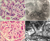 | Figure 1Pathologic findings of renal biopsies. (A) First biopsy: The glomerulus shows marked hypercellularity due to mesangial and endocapillary proliferation, and inflammatory cell infiltration. Hyaline thrombi are clearly observed in capillary lumen (H&E, ×400). (B) First biopsy: Electron microscopy image shows electron dense deposits at mesangium and subendothelial layer. (C) Second biopsy: No significant mesangial matrix widening, mesangial cell proliferation and endocapillary wall thickness are shown (H&E, ×400). (D) Second biopsy: Electron dense deposits are observed at mesangium and subepithelial locations along the glomerular basement membranes by electron microscopy.
|
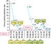 | Figure 2Changes of proteinuria according to treatment with cyclophosphamide and mycophenolate mofetil. The renal rebiopsy was performed and proteinuria was improved significantly after 3 cycles of cyclophosphamide and mycophenolate mofetil administration. CYP: cyclophosphamide, MMF: mycophenolate mofetil.
|
References
1. Bihl GR, Petri M, Fine DM. Kidney biopsy in lupus nephritis: look before you leap. Nephrol Dial Transplant. 2006. 21:1749–1752.
2. Seshan SV, Jennette JC. Renal disease in systemic lupus erythematosus with emphasis on classification of lupus glomerulonephritis: advances and implications. Arch Pathol Lab Med. 2009. 133:233–248.
3. Fiehn C. Early diagnosis and treatment in lupus nephritis: how we can influence the risk for terminal renal failure. J Rheumatol. 2006. 33:1464–1466.
4. Faurschou M, Starklint H, Halberg P, Jacobsen S. Prognostic factors in lupus nephritis: diagnostic and therapeutic delay increases the risk of terminal renal failure. J Rheumatol. 2006. 33:1563–1569.
5. Weening JJ, D'Agati VD, Schwartz MM, Seshan SV, Alpers CE, Appel GB, et al. The classification of glomerulonephritis in systemic lupus erythematosus revisited. J Am Soc Nephrol. 2004. 15:241–250.
6. Christopher-Stine L, Siedner M, Lin J, Haas M, Parekh H, Petri M, et al. Renal biopsy in lupus patients with low levels of proteinuria. J Rheumatol. 2007. 34:332–335.
7. Sidiropoulos PI, Kritikos HD, Boumpas DT. Lupus nephritis flares. Lupus. 2005. 14:49–52.
8. Moroni G, Pasquali S, Quaglini S, Banfi G, Casanova S, Maccario M, et al. Clinical and prognostic value of serial renal biopsies in lupus nephritis. Am J Kidney Dis. 1999. 34:530–539.
9. Daleboudt GM, Bajema IM, Goemaere NN, van Laar JM, Bruijn JA, Berger SP. The clinical relevance of a repeat biopsy in lupus nephritis flares. Nephrol Dial Transplant. 2009. 24:3712–3717.
10. Alsuwaida A, Husain S, Alghonaim M, Aloudah N, Alwakeel J, Ullah A, et al. Strategy for second kidney biopsy in patients with lupus nephritis. Nephrol Dial Transplant. 2012. 27:1472–1478.
11. Eiro M, Katoh T, Watanabe T. Risk factors for bleeding complications in percutaneous renal biopsy. Clin Exp Nephrol. 2005. 9:40–45.
12. Wiseman DA, Hawkins R, Numerow LM, Taub KJ. Percutaneous renal biopsy utilizing real time, ultrasonic guidance and a semiautomated biopsy device. Kidney Int. 1990. 38:347–349.
13. Whittier WL, Korbet SM. Timing of complications in percutaneous renal biopsy. J Am Soc Nephrol. 2004. 15:142–147.
14. Esdaile JM, Joseph L, MacKenzie T, Kashgarian M, Hayslett JP. The pathogenesis and prognosis of lupus nephritis: information from repeat renal biopsy. Semin Arthritis Rheum. 1993. 23:135–148.
15. Lightstone L. Lupus nephritis: where are we now? Curr Opin Rheumatol. 2010. 22:252–256.
 XML Download