

 PDF
PDF ePub
ePub Citation
 Print
Print


Introduction
Systemic sclerosis (SSc) is a systemic autoimmune or connective tissue disease of unknown etiology, which is characterized by fibrosis of the skin and internal organs as well as widespread vascular involvement. Among these features, vasculopathy has been accepted as an important and primary process in SSc (1,2). The mechanism of vasculopathy in SSc has not yet been fully elucidated, but a disrupted or inappropriate repair process following endothelial damage seems to result in vasculopathy (3). Although microvascular damage has been well described as a major cause of SSc, recent reports have demonstrated an increased prevalence of macrovascular disease and atherosclerosis as well (4-6).
Metabolic syndrome (MetS), also known as syndrome X or the insulin resistance syndrome, is a constellation of metabolic disturbances such as abdominal obesity, atherogenic dyslipidemia, hypertension (HTN), disturbed glucose metabolism, and insulin resistance, all of which are independent risk factors for atherosclerosis and cardiovascular diseases (CVDs) (7,8). When grouped together, these risk factors are associated with cardiovascular risks beyond the sum of its individual components (9). Although several diagnostic criteria for MetS have been established to date, no consensus has been reached regarding the definition of MetS. However, the Third report of the National Cholesterol Education Program's Adult Treatment Panel (NCEP-ATP III) 2004 (10) and the International Diabetes Federation (IDF) 2005 (11) definition for MetS have been most widely used due to simplicity for clinical use (12). Previous studies have reported a higher prevalence of MetS in other rheumatic diseases, such as rheumatoid arthritis (RA) and systemic lupus erythematosus (SLE) (7,8). However, no direct studies have investigated the prevalence of MetS in patients with SSc as compared with healthy subjects (13) despite an increased risk for atherosclerosis and CVDs in SSc.
Insulin resistance is considered a key pathogenic factor for MetS and may explain the interaction between chronic inflammatory diseases and CVDs (7). Various methods have been employed for measuring insulin resistance to date. Although the hyperinsulinemic euglycemic glucose clamp method is considered the gold standard, it is both labor and time consuming (14). Homeostatic assessment model-insulin resistance (HOMA-IR), which is simply calculated from fasting blood insulin and glucose concentrations, closely correlates with the hyperinsulinemic euglycemic glucose clamp method and is useful index of insulin resistance, especially in clinical settings (14). To the best of our knowledge, little is known about insulin resistance as measured by HOMA-IR in SSc patients, unlike other rheumatic diseases, such as RA and SLE.
In this study, we evaluated the frequency of MetS and the magnitude of insulin resistance measured by HOMA-IR in female patients with SSc in comparison with those of healthy subjects and we also investigated risk factors associated with HOMA-IR and the presence of MetS in female patients with SSc.
Materials and Methods
Study designs and subjects
In a cross-sectional setting, our study included 35 consecutive patients with SSc and 84 age- and sex-matched healthy subjects aged from 20 to 70 years, who were recruited from a single regional rheumatism center of a tertiary hospital in South Korea between 2009 and 2010. Due to the limited number of male patients with SSc in the center, only female SSc patients were included in the study. All patients fulfilled the preliminary classification criteria of the American College of Rheumatology (15). The following patients were excluded from the study: 1) patients with rheumatic diseases other than SSc; 2) patients who had previous CVDs including ischemic heart disease and stroke; and 3) patients who refused to participate in this study. Healthy subjects were selected randomly from applicants for an annual health check in the same center and had no history of rheumatic or CVDs. Patients with SSc and healthy subjects were frequency matched for age (±1 year) and all subjects were South Korean. Written informed consent based on the Helsinki Declaration was obtained from each subject. This study was approved by the Research and Ethical Review Board of Pusan National University Hospital, Busan, South Korea.
Assessments
Weights and heights of all participants were measured by portable calibrated electronic weighting scales and portable inflexible measuring bars, respectively. Body mass index (BMI) was calculated by dividing body weight by the square of height in meters (kg/m2). Waist circumference was measured at the end of normal expiration, with arms relaxed at the sides, at the midpoint between the lower part of the lowest rib and the highest point of the superior iliac crest on the mid-axillary line, using constant tension tape. Blood pressure was determined, using a TM-2655P apparatus (A&D Company Ltd, Tokyo, Japan), as the average of 2 measurements which were obtained 5 minutes apart after participants rested for at least 10 minutes. HTN was defined as blood pressure ≥140/90 mmHg or requiring antihypertensive medications.
Blood samples were taken between 8:00 AM and 10:00 AM after 10 hour of overnight fasting. Blood test included: fasting glucose and insulin, fasting lipid profile (total cholesterol (TC), triglyceride (TG), high density lipoprotein (HDL) cholesterol, low density lipoprotein (LDL) cholesterol), and high-sensitivity C-reactive protein (hsCRP). Blood concentrations of insulin were determined using human radioimmunoassay (Coat-A-Count® human radioimmunoassay, Siemens Healthcare Diagnostics, USA). TC, HDL cholesterol, and TG concentrations were measured using of an enzymatic colorimetric reagent (Roche Diagnostics, Switzerland) on a P-800 Modular (Roche, Switzerland). LDL cholesterol was calculated by using the Fridewald formula. The concentrations of hsCRP were analyzed using a particle-enhanced immunoturbidimetric assay (Tina-quant C-reactive protein, Roche Diagnostics, Switzerland) according to the manufacturer's instruction on the automated analyzer, a P-800 Modular (Roche, Switzerland).
Patients with SSc were classified as having either limited SSc or diffuse SSc based on the extent of skin involvement according to the LeRoy et al. (16). For all SSc patients, pharmacy and medical records were reviewed, and the cumulative glucocorticoids (GCs) dose was calculated by multiplying the current daily dose by the number of days for which patients had received GCs since they were first prescribed. For the examination of organ involvements, following procedures were performed: The diagnosis of interstitial lung disease (ILD) was based on high resolution computed tomography (HRCT); Pulmonary arterial hypertension (PAH) was defined as pulmonary arterial pressure (PAP) >35 mmHg on at least 2 occasions, as measured by color Doppler echocardiography; Gastrointestinal tract involvement was determined by clinical symptoms, including dysphagia, reflux esophagitis requiring the use of proton pump inhibitors, or small bowel bacterial overgrowth. Skin involvement was assessed using the modified Rodnan skin score measured by a rheumatologist. The following autoantibody profiles were also measured: antinuclear antibody (ANA) and anticentromere antibody (indirect immunofluorescence on Hep-2 cells, cut-off value: 1:40) and anti-SCL-70 (immunoblot testing, anti-ENA profile class-1 Euroline).
Measurement of insulin resistance and definition of MetS
Insulin resistance was evaluated by homeostasis model assessment for insulin resistance (HOMA-IR) which was calculated with the formula defined by Matthews et al. (17) as follows: HOMA-IR=[fasting serum insulin (µIU/mL)×fasting serum glucose (mg/dL)×0.055÷22.5].
MetS was defined according to the NCEP-ATP III 2004 (10), using the Asian criteria for central obesity (18) when 3 or more of the following components were present: 1) increased waist circumference to ≥90 cm in men or ≥80 cm in women; 2) elevated blood pressure to ≥130/85 mmHg or requiring drug therapy; 3) elevated serum TG level to ≥150 mg/dL; 4) reduced serum HDL-cholesterol to ≤40 mg/dL in men or ≤50 mg/dL in women; and 5) elevated fasting glucose level to ≥100 mg/dL or requiring drug therapy.
Statistical analysis
No formal sample size calculation was conducted. Data were presented as mean±standard deviation, median (inter-quartile range), or number (percentage), as appropriate. Comparisons between SSc patients and healthy controls were performed using the 2-tailed Student's t test or the Mann-Whiteny U test for continuous variables and the χ2 test or the Fisher's exact test for categorical variables, as appropriate. Possible associated factors with HOMA-IR in female SSc patients were investigated using the Spearman's correlation analysis. Statistical significance of predictors for the presence of MetS in patients with SSc was tested by the univariable logistic regression analysis. Due to the small sample size of SSc patients and number of subjects with MetS, we could not carry out the multivariable logistic regression analysis considering that the precision of the logistic regression model becomes problematic when the ratio of the number of events per independent variable becomes small (19). All statistical analyses were performed using STATA 11.1 for windows (StataCorp LP, College Station, TX, USA).
Results
Characteristics of patients with SSc
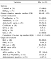
Table 1 shows clinical characteristic of 35 female patients with SSc. Of SSc patients, 17 had limited SSc and 18 had diffuse SSc. Twenty-one patients (60%) were taking GCs and median cumulative GCs dose was 1126.3 mg. Twenty-seven patients (77.1%) were receiving vasodilators; 2 (5.7%) had a history of HTN. The majority of patients (80%) had ILD, whereas 5 patients (14.3%) had PAH.
Metabolic perturbations of patients with SSc and health subjects
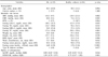
Table 2 shows demographics and metabolic risk factors of study population. No significant differences were seen between the two groups according to age, the percentage of current smoker, diastolic blood pressure (DBP), fasting LDL and HDL cholesterol, height, and the proportion of subjects with HTN and type II diabetes mellitus. Systolic blood pressure (SBP), weight, BMI, waist circumference, and fasting glucose concentration were significantly lower in patients with SSc than in healthy controls, whereas SSc patients had significantly higher TG, fasting insulin concentrations, and hsCRP. Interestingly, there was no significant difference in the prevalence of MetS between the two groups (p=0.425), whereas HOMA-IR in patients with SSc was significantly higher than in healthy subjects (p=0.001).
Table 3 demonstrates the frequency of individual MetS features in the two groups. In comparison with healthy subjects, significantly more patients with SSc had increased TG concentrations (p=0.004). Less SSc patients tended to meet the waist circumference criterion of the Asian criteria for central obesity, but the difference was not statistically significant (p=0.051). There were no significant differences between the two groups in BP, HDL-cholesterol, and blood glucose concentrations criteria.
Risk factors associated with MetS and HOMA-IR in patients with SSc
Table 4 shows the factors associated with MetS in 35 female patients with SSc. Higher hsCRP and longer disease duration tended to correlated with the presence of MetS, but did not reach statistical significance (p=0.069 and 0.062, respectively). Age, BMI, HOMA-IR, cumulative GCs dose, MRSS, or subtype of SSc did not show significant relationships with MetS in patients with SSc. As mentioned above, multivariable analyses were not performed due to the small number of SSc patients. In addition, there were no significant differences in age, hsCRP, BMI, HOMA-IR, disease duration, cumulative GCs dose, MRSS or the proportion of limited SSc cases according to the presence or absence of MetS in SSC patients (data not shown).
HOMA-IR in patients with SSc was positively correlated with BMI (ρ=0.454, p=0.007) and waist circumference (ρ=0.434, p=0.012). Age, SBP, DBP, fasting LDL cholesterol, TG, and HDL cholesterol, hsCRP, disease duration, and cumulative GCs dose were not significantly correlated with HOMA-IR in SSc patients (data not shown). SSc patients with positive anti-SCL-70 have a significantly lower HOMA-IR than those without anti-SCL-70 (0.78±0.32 vs 1.70±0.93, p=0.001), but there was no significant difference in HOMA-IR between patients with and without anticentromere antibody (p=0.595). In addition, HOMA-IR did not differ significantly between patients with limited SSc and those with diffuse SSc (p=0.235).
Discussion
To our knowledge, this is the first study that evaluated the prevalence of MetS and the magnitude of insulin resistance measured by HOMA-IR in Asian patients with SSc. In our study, there was no significant difference between in the frequency of MetS between patients with SSc and healthy subjects, whereas patients with SSc had a significantly higher HOMA-IR than healthy controls. In addition, BMI and waist circumference were positively correlated with HOMA-IR in patients with SSc. Although hsCRP and disease duration were likely to be associated with the presence of MetS, no significant predictor for SSc patients with MetS were observed in our study.
Over the last decade, many studies have demonstrated the prevalence of MetS in rheumatic diseases such as RA (20-26), SLE (27-30), ankylosing spondylitis (AS) (31), and psoriatic arthritis (PsA) (32). The prevalence of MetS in patients with rheumatic diseases ranged from 19.6% to 58.1% in previous reports. Various factors such as ethnicity, geographic area, life style, age, sex, the definition of MetS, and the type of rheumatic diseases might have led to the diversity in the frequency of MetS in rheumatic diseases. However, the prevalence of MetS were consistently higher in patients with SLE, AS, and PsA than in healthy controls except in a study by Sabio et al. (29) which demonstrated only an insignificantly increased prevalence of MetS in SLE patients as compared with healthy subjects (p=0.083). Although whether the prevalence of MetS in RA patients is higher than the general population was somewhat compelling in previous reports (20-26), it has generally been considered that MetS is more prevalent among patients with RA (7,8). The majority of studies support the observation that rheumatic diseases are closely associated with an increased prevalence of MetS. Our study is noteworthy, however, because we found that that the prevalence of MetS in SSc patients was not significantly higher than in healthy subjects.
MetS is considered a proinflammatory state and closely associated with low grade systemic inflammation (33). In addition, a rise in adipokines and proinflammatory cytokines including tumor necrosis factor α (TNFα) and interleukin-6 (IL-6) can promote insulin resistance and thus increase the prevalence of MetS (8). Therefore, inflammatory biomarkers are frequently elevated in individuals with MetS and, conversely, the prevalence of MetS is higher in patients with chronic inflammatory statuses such as rheumatic diseases (8). Taken together, inflammation can provide a plausible explanation for the link between MetS and rheumatic diseases. However, no statistical difference in the prevalence of MetS between patients with SSc and healthy controls in our study is inconsistent with previous findings. We conjecture that the magnitude of inflammation in SSc patients might not be as high as in patients with other rheumatic diseases such as RA, and also inflammation might interact with the occurrence of MetS differently according to rheumatic diseases. The complex interplay between inflammation and MetS in chronic inflammatory or rheumatic diseases is not fully understood. Thus, further study is needed to confirm the exact mechanism by which inflammatory status can increase the occurrence of MetS in various rheumatic diseases.
Long term use of GCs, which are widely used to control various rheumatic diseases, is known to cause metabolic disturbances including HTN, dyslipidemia and diabetes. However, there is conflicting evidence for the association between GCs use and MetS in patients with various rheumatic diseases. Except for a study by Bultink et al. (28) which showed that intravenous methylprednisolone use was positvely associated with MetS score in SLE patients, most studies have not demonstrated a causal relationship between GCs use and MetS in patients with rheumatic disease. Similarly, in our patients, cumulative GCs dose was not associated with the frequency of MetS in SSc patients in our study. In addition, although patients with AS are likely to require lower-dose and shorter-duration of GCs therapy than those with RA and SLE, the prevalence of MetS is higher in AS patients than in healthy controls as with other rheuamtic diseases (31). Taken together, the role of GCs in the occurrence of MetS has not yet been determined.
In our study, the degree of insulin resistance as measured by HOMA-IR was significantly higher in patients with SSc than in healthy subject although there was no difference in the prevalence of MetS between the two groups. Considering that insulin resistance is the key pathophysiologic factor for MetS, our findings seemed to be unanticipated. A possible explanation for these results may be that, while insulin resistance is an important contributor to MetS, insulin resistance may not be sufficient, by itself, for the occurrence of MetS (34). This concept was corroborated by the report of Hanely et al. (35), which demonstrated that the components of MetS can largely be grouped by two separate factors, metabolic and blood pressure, using factor analysis. In light of these findings, the higher HOMA-IR in our patients with SSc may not correlate with the occurrence of MetS. Similarly, La Montagna et al. (20) also showed no difference in the occurrence in MetS between RA patients and healty controls despite a signficantly higher HOMA-IR values in RA patients as compared with healthy controls (p<0.001). The mechanism of increased insulin resistacne as measured by HOMA-IR in SSc patients is not fully understood. We assumed that tissue growth factor-β (TGF-β), an important soluble mediator in SSc (3), may play a role in increased insulin restistance (36). Additionally, a higer inflammation degrees in SSc patients could contribute to the increased HOMA-IR.
Obesity in patients with SSc has not yet been systematically investigated. In this study, BMI and waist circumference were significantly lower in patients with SSc than in healthy subjects. In line with our findings, Borba et al. (2) and Mok et al. (37) also reported less obesity in SSc patients as compared with healthy subjects. Although why patients with SSc were found to have less obesity in our study and previous studies is not fully elucidated, these findings might affect the lack of prevalence of MetS in SSc patient compared with healthy subjects in our study considering that abdominal obesity has been recognized as a major contributor to insulin resistance and MetS pathophysiology. However, Hettema et al. (38) did not confirm less obesity in SSc patients. Further researches are needed to delineate the relationship between obesity and SSc.
Some differences in traditional risk factors between patients with SSc and healthy subjects emerged in this study. Firstly, SSc patients had a significantly lower SBP than healthy subjects (p=0.002). This result was partly owing to the use of vasodilators (indicated for HTN and Raynaud's phenomenon), which were given to the majority of SSc patients (77.1%). Low blood pressure can be measured in normotensive SSc patients taking vasodilators for Raynaud's phenomenon. Meanwhile, no significant differences in the proportion of HTN between SSc patients and healthy subjects were observed in our study. Similar results were observed in the study by Mok et al (37). However, since blood pressure measurements in SSc patients taking vasodilators should be interpreted cautiously, whether SSc patients have a lower blood pressure is yet to be determined. Secondly, TG concentrations were higher in patients with SSc than in healthy controls (p=0.012). However, the lipid profiles of SSc patients in previous studies were not always consistent with those in our study (2,38). A distinctive pattern of lipid profile in SSc patients has not yet been validated.
We note a number of potential limitations to our study. First, due to the small number of SSc patients and the small percentage of subjects with MetS among these patients, only female patients were included and multivariable analyses to investigate risk factors for the presence of MetS were not carried out in the present study. Thus, the causality between clinical makers and MetS in SSc patents was not fully evaluated. Second, only the subjects who visited a single tertiary center were included only in the present study, which could lead to a selection bias. Thus, study population in our study might be different from the general population and SSc patients from other centers. Third, in our study, the frequency of MetS in healthy subjects (14.3%) was relatively low, as compared with data from a nationwide survey using the NCEP-ATPIII 2001 criteria (38.7) (39). Apart from the difference in the definition of MetS, we speculate that this discrepancy in the prevalence of MetS might be due to the characteristics of the geographic area in our study. The majority of subjects in this study were residing in coastal areas and the distinctive lifestyle may exist and affect the prevalence of MetS. Actually, Kang et al. (40) elegantly reported that the frequency of MetS in 2519 female healthy subjects from the same center of our study was 15.6% according to the NCEP-ATPIII 2004 criteria, which is similar to ours (40). Additionally, our study did not include the physical activity of study subjects which can affect obesity and the presence of MetS.
To summarize, our study showed that the frequency of MetS did not differ significantly between female SSc patients and healthy controls. Meanwhile, HOMA-IR and TG in SSc patients is higher than in healthy controls, which suggest that clinicians need to pay special attention to increased insulin resistance and a deleterious lipoprotein pattern in SSc patients.
Conclusion
In this preliminary study, the frequency of MetS was not significantly higher in patients with SS than in healthy subjects, whereas SSc patients had increased insulin resistance as measured by HOMA-IR. Since the prevalence of MetS is higher in other rheumatic diseases such as RA, SLE, AS, and PsA than in the general population, our results may be noteworthy despite the small sample size.


 XML Download