Abstract
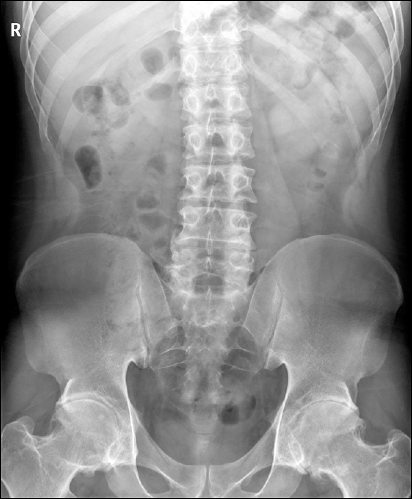
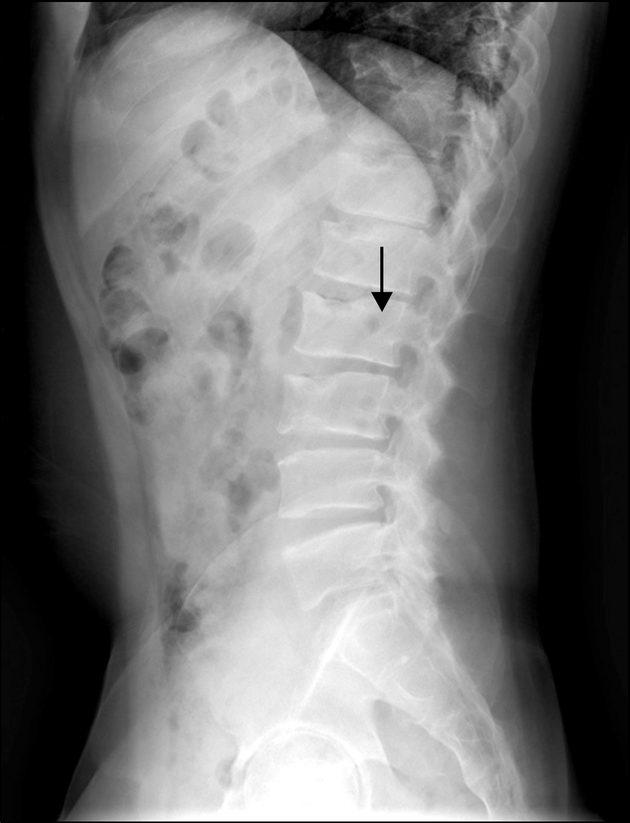
The spondyloepiphyseal dysplasia tarda (SEDT) is a hereditary arthropathy that progressively leads to deform-ities of small and large joints, irregularities of the end plates of vertebral bodies, which causes joint restriction, short stature, and gait difficulties. The typical radio-graphic findings of SEDT are generalized platyspondyly and dysplasia of the epiphyses, resulting in premature arthrosis. Clinically SEDT is manifested as a form of short-trunk dwarfism and early arthrosis in the period from late childhood to adolescence. The major clinical importance of this rare disease is similarity to juvenile idiopathic arthritis (JIA), which has a rather different prognosis and treatment. A few cases of SEDT have been published. However, no cases have been reported in South Korea. We describe the case of a 29-year old man who suffered from back and multiple joint pain, who was misdiagnosed as having ankylosing spondylitis. We evaluated the patient clinically and radiographically in greater detail, and changed his diagnosis to SED tarda.
Go to : 
References
1. Maroteaux P, Lamy M, Bernard J. La dysplasie spondy-lo-epiphysaire tardive. Presse Med. 1957; 65:1205–8.
2. Gupta R, Gulati N, Hatimota P, Gulati M, Kumar A. Spondyloepiphyeal dysplasia tarda mimicking juvenile idiopathic arthritis-a case report. J Indian Rheumatol Assoc. 2002; 10:22–3.
3. Bal S, Kocyigit H, Turan Y, Gurgan A, Bayram KB, Güvenc A, et al. Spondyloepiphyseal dysplasia tarda: four cases from two families. Rheumatol Int. 2009; 29:699–702.

4. Wynne-Davies R, Gormley J. The prevalence of skeletal dysplasias. An estimate of their minimum frequency and the number of patients requiring orthopaedic care. J Bone Joint Surg Br. 1985; 67:133–7.

5. Carter CO, Sutcliffe J. Genetic varieties of spondyloepiphyseal dysplasia. Jellife AM, Strickland B, editors. Symposium ossium. p. 218–24. Edinburgh London: Livingstone;1970.
6. Kocyigit H, Arkun R, Ozkinay F, Cogulu O, Hizli N, Memis A. Spondyloepiphyseal dysplasia tarda with progressive arthropathy. Clin Rheumatol. 2000; 19:238–41.

7. Leroy JG, Leroy BP, Emmery LV, Messiaen L, Spranger JW. A new type of autosomal recessive spondyloepiphyseal dysplasia tarda. Am J Med Genet A. 2004; 125A:49–56.

8. Langer LO JR. Spondyloepiphysial dysplasia tarda. Hereditary chondrodysplasia with characteristic vertebral configuration in the adult. Radiology. 1964; 82:833–9.
9. Arslanoglu S, Murat H, Ferah G. Spondyloepiphyseal dysplasia tarda with progressive arthropathy: an important form of osteodysplasia in the differential diagnosis of juvenile rheumatoid arthritis. Pediatr Int. 2000; 42:561–3.

10. Spranger J, Albert C, Schilling F, Bartsocas C. Progressive pseudorheumatoid arthropathy of childhood (PPAC): a hereditary disorder simulating juvenile rheumatoid arthritis. Am J Med Genet. 1983; 14:399–401.

Go to : 


 PDF
PDF ePub
ePub Citation
 Print
Print


 XML Download