References
1. Johnso TR, Weckbach S, Kellner H, Reiser MF, Becker CR. Clinical image: Dual-energy computed tomographic molecular imaging of gout. Arthritis Rheum. 2007; 56:2809.

2. Nicolaou S, Yong-Hing CJ, Galea-Soler S, Hou DJ, Louis L, Munk P. Dual-energy CT as a potential new diagnostic tool in the management of gout in the acute setting. AJR Am J Roentgenol. 2010; 194:1072–8.

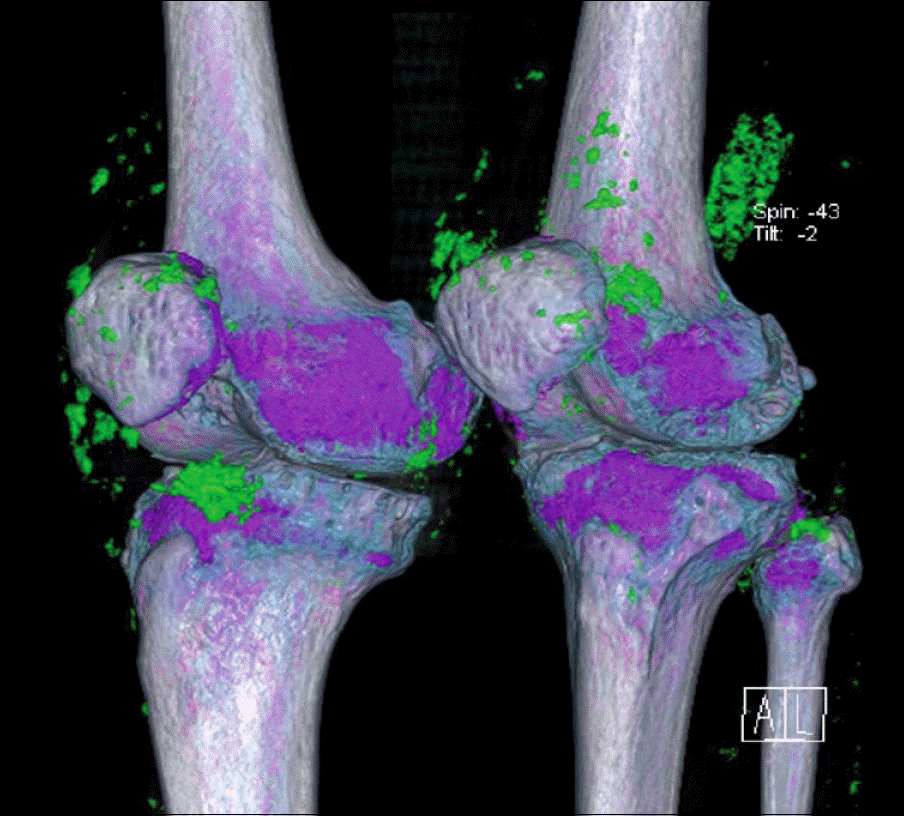
Figure 1.
Volumn-rendered three-dimensional dual-energy CT (DECT) image shows multiple monosodium urate deposits (i.e., tophi), which are responsible for this patient's symptoms as seen in both knees.

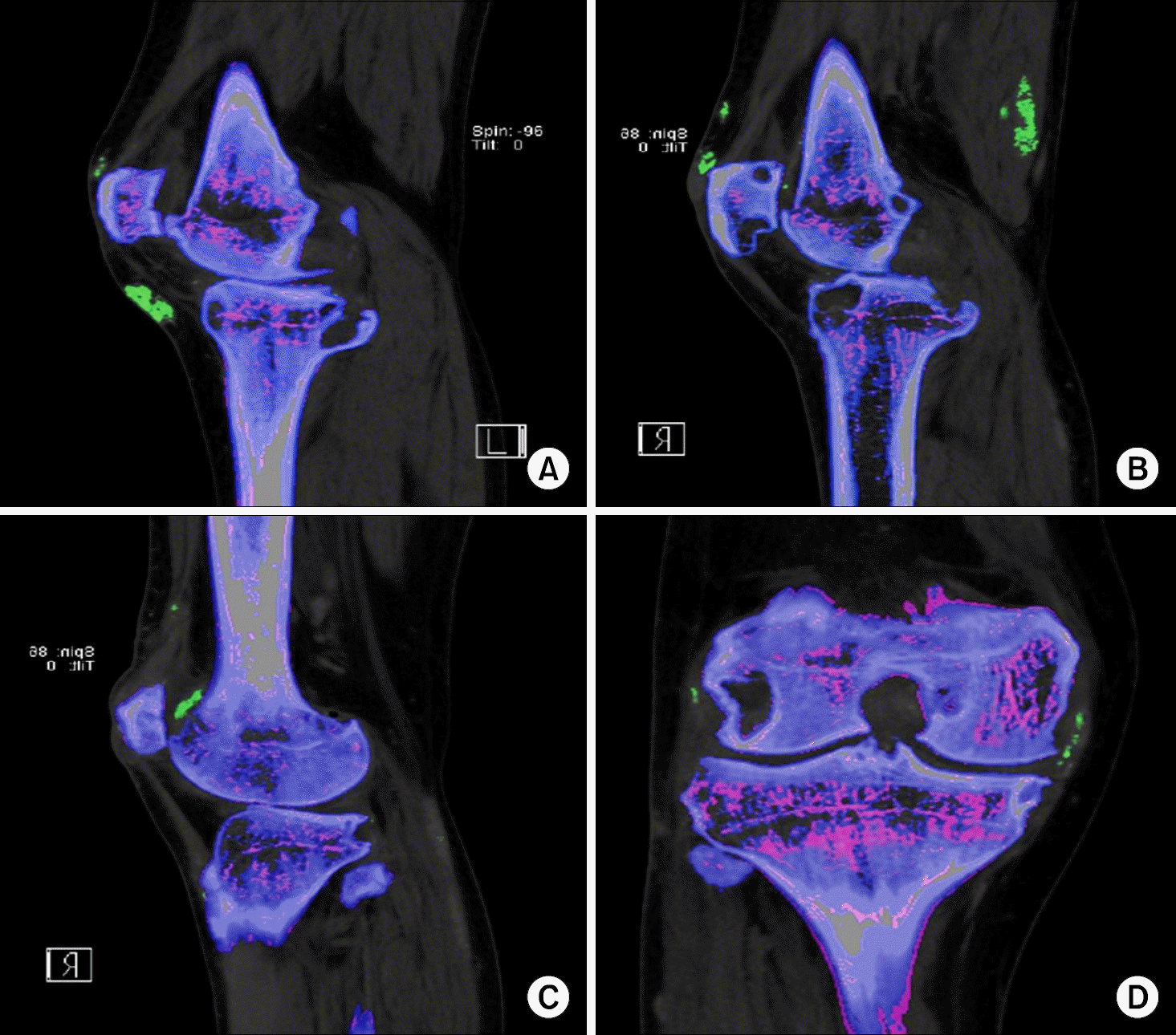
Figure 2.
Dual energy CT (DE-CT) images of uric acid deposits in various anatomical locations in both knees of the patent: (A) Lt Infrapatellar bursa, (B) Rt prepa-tellar bursa and semi-membrane-ous muscle, (C) Rt suprapatellar bursa, (D) Rt medial collateral ligament. With the application of the three material decomposition algorithm, uric acid deposits are depicted in green, whereas calc-ium in bone is depicted in blue. Multiple large subchondral and paraarticular bone erosions of both knee joints with severe joint space narrowing are found in each image.


 PDF
PDF ePub
ePub Citation
 Print
Print


 XML Download