Journal List > J Rheum Dis > v.18(1) > 1063910

Park, Park, Min, and Kim: Risk Factors for Low Bone Mineral Density in Korean Patients with Systemic Lupus Erythematosus

Abstract

Objective

To determine the degree and risk factors for decreased bone mineral density (BMD) in patients with systemic lupus erythematosus (SLE).

Methods

One hundred and one patients with SLE and 57 age- and gender-matched healthy controls were enrolled in this study. The BMD was measured by dual energy X-ray absorptiometry (DXA). The laboratory findings and clinical variables evaluated in the SLE patients consisted of disease duration, SLE disease activity index (SLEDAI), and medications, including mean and cumulative dose of glucocorticoid. At the time of the clinical and laboratory assessment, the levels of serum osteocalcin, serum FSH/LH, urine deoxypyridinoline (DPD), and serum cytokines, such as IL-6 and soluble receptor activator of NF-kB ligand (RANKL), were determined in SLE patients using a enzyme-linked immunosorbent assay.

Results

The BMD T score decreased in patients with SLE compared to the healthy controls (−1.11 versus −0.41, p=0.001 at lumbar spine, −0.84 versus −0.01, p<0.001 at femur neck, −1.20 versus −0.45, p<0.001 at total hip, respectively). Osteoporosis and osteopenia was present in 16.8% and 46.5% of patients, respectively. Multiple regression analysis revealed a low BMD in the lumbar spine to be associated with increased FSH, low BMI and cumulative glucocorticoid dose. A low BMD in the hip and femur neck was associated with increased FSH, low BMI, and duration of glucocorticoid. On the other hand, the levels of osteocalcin, deoxypyridinoline (DPD), IL-6, and soluble RANKL were similar in patients with a low BMD and those with normal BMD.

Conclusion

Osteoporosis and osteopenia are more common in young Korean SLE patients than in control subjects. Elevated FSH, low BMI, and the use of glucocorticoid are independent risk factors linked to a decreased BMD in Korean patients with SLE.

References

1. Urowitz MB, Bookman AA, Koehler BE, Gordon DA, Smythe HA, Ogryzlo MA. The bimodal mortality pattern of systemic lupus erythematosus. Am J Med. 1976; 60:221–5.
2. Ginzler EM, Diamond HS, Weiner M, Schlesinger M, Fries JF, Wasner C, et al. A multicenter study of outcome in systemic lupus erythematosus. I. Entry variables as predictors of prognosis. Arthritis Rheum. 1982; 25:601–11.
3. Ward MM, Pyun E, Studenski S. Mortality risks associated with specific clinical manifestations of systemic lupus erythematosus. Arch Intern Med. 1996; 156:1337–44.
4. Ward MM, Pyun E, Studenski S. Causes of death in systemic lupus erythematosus. Longterm followup of an inception cohort. Arthritis Rheum. 1995; 38:1492–9.
5. Trager J, Ward MM. Mortality and causes of death in systemic lupus erythematosus. Curr Opin Rheumatol. 2001; 13:345–51.
6. Gordon C. Longterm complications of systemic lupus erythematosus. Rheumatology (Oxford). 2002; 41:1095–100.
7. Christian JC, Yu PL, Slemenda CW, Johnston CC Jr. Heritabili-ty of bone mass: a longitudinal study in aging male twins. Am J Hum Genet. 1989; 44:429–33.
8. Pocock NA, Eisman JA, Hopper JL, Yeates MG, Sambrook PN, Eberl S. Genetic determinants of bone mass in adults. A twin study. J Clin Invest. 1987; 80:706–10.
9. Johnston CC Jr, Miller JZ, Slemenda CW, Reister TK, Hui S, Christian JC, et al. Calcium supplementation and increases in bone mineral density in children. N Engl J Med. 1992; 327:82–7.
10. Morrison NA, Qi JC, Tokita A, Kelly PJ, Crofts L, Nguyen TV, et al. Prediction of bone density from vitamin D receptor alleles. Nature. 1994; 367:284–7.
11. Dalsky GP. The role of exercise in the prevention of osteoporosis. Compr Ther. 1989; 15:30–7.
12. Pacifici R. Estrogen, cytokines, and pathogenesis of postmenopausal osteoporosis. J Bone Miner Res. 1996; 11:1043–51.
13. Anderson GL, Limacher M, Assaf AR, Bassford T, Beresford SA, Black H, et al. Effects of conjugated equine estrogen in postmenopausal women with hysterectomy: the Women's Health Initiative randomized controlled trial. JAMA. 2004; 291:1701–12.
14. Canalis E. Clinical review 83: Mechanisms of glucocorticoid action in bone: implications to glucocorticoid-induced osteoporosis. J Clin Endocrinol Metab. 1996; 81:3441–7.
15. Haugeberg G, Ørstavik RE, Kvien TK. Effects of rheumatoid arthritis on bone. Curr Opin Rheumatol. 2003; 15:469–75.
16. Lacativa PG, Farias ML. Osteoporosis and inflammation. Arq Bras Endocrinol Metabol. 2010; 54:123–32.
17. Tanaka Y, Watanabe K, Suzuki M, Saito K, Oda S, Suzuki H, et al. Spontaneous production of bone-resorbing lymphokines by B cells in patients with systemic lupus erythematosus. J Clin Immunol. 1989; 9:415–20.
18. MacDonald BR, Gowen M. Cytokines and bone. Br J Rheumatol. 1992; 31:149–55.
19. Report of a WHO Study Group. Assessment of fracture risk and its application to screening for postmenopausal osteoporosis. World Health Organ Tech Rep Ser. 1994; 843:1–129.
20. Redlich K, Ziegler S, Kiener HP, Spitzauer S, Stohlawetz P, Bernecker P, et al. Bone mineral density and biochemical param-eters of bone metabolism in female patients with systemic lupus erythematosus. Ann Rheum Dis. 2000; 59:308–10.
21. García-Carrasco M, Mendoza-Pinto C, Escárcega RO, Jiménez-Hernández M, Etchegaray Morales I, Munguía Realpozo P, et al. Osteoporosis in patients with systemic lupus erythematosus. Isr Med Assoc J. 2009; 11:486–91.
22. Kipen Y, Buchbinder R, Forbes A, Strauss B, Littlejohn G, Morand E. Prevalence of reduced bone mineral density in systemic lupus erythematosus and the role of steroids. J Rheumatol. 1997; 24:1922–9.
23. Bhattoa HP, Bettembuk P, Balogh A, Szegedi G, Kiss E. Bone mineral density in women with systemic lupus erythematosus. Clin Rheumatol. 2002; 21:135–41.
24. Bhattoa HP, Kiss E, Bettembuk P, Balogh A. Bone mineral density, biochemical markers of bone turnover, and hormonal status in men with systemic lupus erythematosus. Rheumatol Int. 2001; 21:97–102.
25. Lakshminarayanan S, Walsh S, Mohanraj M, Rothfield N. Factors associated with low bone mineral density in female patients with systemic lupus erythematosus. J Rheumatol. 2001; 28:102–8.
26. Tan EM, Cohen AS, Fries JF, Masi AT, McShane DJ, Rothfield NF, et al. The 1982 revised criteria for the classification of systemic lupus erythematosus. Arthritis Rheum. 1982; 25:1271–7.
27. Park HA, Park JK, Park SA, Lee JS. Age, menopause, and cardiovascular risk factors among korean middle-aged women: the 2005 Korea National Health and Nutrition Examination Survey. J Womens Health (Larchmt). 2010; 19:869–76.
28. Bombardier C, Gladman DD, Urowitz MB, Caron D, Chang CH. Derivation of the SLEDAI. A disease activity index for lupus patients. The Committee on Prognosis Studies in SLE. Arthritis Rheum. 1992; 35:630–40.
29. Genant HK, Cooper C, Poor G, Reid I, Ehrlich G, Kanis J, et al. Interim report and recommendations of the World Health Organization Task-Force for Osteoporosis. Osteoporos Int. 1999; 10:259–64.
30. Molina B, Lassaletta A, Andion M, Gonzalez-Vicent M, López-Pino MA, et al. A Persistent epidural mass in a child with B-lin-eage ALL. Pediatr Blood Cancer. 2010; 55:727–9.
31. Warne GL, Fairley KF, Hobbs JB, Martin FI. Cyclophospha-mide-induced ovarian failure. N Engl J Med. 1973; 289:1159–62.
32. Mok CC, Lau CS, Wong RW. Risk factors for ovarian failure in patients with systemic lupus erythematosus receiving cyclophosphamide therapy. Arthritis Rheum. 1998; 41:831–7.
33. Hofbauer LC, Gori F, Riggs BL, Lacey DL, Dunstan CR, Spelsberg TC, et al. Stimulation of osteoprotegerin ligand and inhibition of osteoprotegerin production by glucocorticoids in human osteoblastic lineage cells: potential paracrine mechanisms of glucocorticoid-induced osteoporosis. Endocrinology. 1999; 140:4382–9.
34. Van Staa TP, Leufkens HG, Cooper C. The epidemiology of cor-ticosteroid-induced osteoporosis: a meta-analysis. Osteoporos Int. 2002; 13:777–87.
35. Dalle Carbonare L, Arlot ME, Chavassieux PM, Roux JP, Portero NR, Meunier PJ. Comparison of trabecular bone micro-architecture and remodeling in glucocorticoid-induced and postmenopausal osteoporosis. J Bone Miner Res. 2001; 16:97–103.
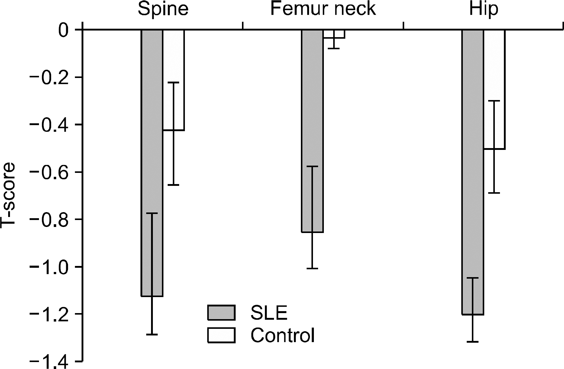
Figure 1.
Bone mineral density at different sites in the patients with SLE and age-and gender-matched healthy controls. The BMD T-score was significantly lower in the SLE patients than in the healthy controls (−1.11±0.13 versus −0.41±0.15, p=0.001 at lumbar spine, −0.84±0.11 versus −0.01±0.15, p<0.001 at femur neck, −1.20±0.11 vs −0.45±0.14, p<0.001 at total hip, respectively). The data is reported as the mean± SEM.
jrd-18-19f1.tif
Table 1.
Characteristics of the SLE patients and age-and gender-matched control subjects
Variables SLE patients (n=101) Control (n=57) p
Age, yr 37.9±12.1 39.8±5.6 0.916
Sex, F (%) 99 (98.0)(%) 55 (96.5) 0.963
Menopausal state, no (%) 25 (24.7) 4 (14.3) 0.001
BMI, kg/m2 21.9±3.0 22.3±3.4 0.553
Dyslipidemia, n (%) 78 (43.1) 30 (35.3) 0.436
Osteocalcin, ng/mL 5.8 9.2 <0.001
  [4.3∼9.2] [7.3∼11.5]  
DPD, nM/mM 45.3 50.3 0.690
  [11.8∼80.1] [23.4∼67.6]  
IL-6, pg/mL 3 [0∼5] 2 [0∼7] 0.921
OPG, pg/mL 1,246 1,042 0.263
  [972.5∼1,557.5] [926∼1,242]  
RANKL, pg/mL 2.5 [1∼5.0] 3 [1.5∼10] 0.182
FSH, IU/L 6.1  
  [3.1∼18.7]    
LH, IU/L 7.9  
  [3.5∼23.3]    
C3, mg/dL 116.9±32.4  
C4, mg/dL 19.6±7.9  
Anti-dsDNA Ab, IU/m L 27.7±5.6  
Hx. of LN, no (%) 20 (19.8)  
HCQ, no (%) 82 (81.2)  
Calcium, no (%) 36 (35.6)  
CYC, no (%) 17 (16.8)  
CsA, no (%) 5 (5)  

The data is reported as mean± SD or median [interquartile range: IQR]. BMI: body mass index, DPD: deoxypyridinoline, IL-6: interleukin-6, OPG: osteoprotegerin, RANKL: receptor activator NF-kB ligand, FSH: follicule stimulating hormone, LH: luteinizing hormone, LN: lupus nephritis, HCQ: hydroxychloroquine, CYC: cyclophosphamide, CsA: cyclosporine A. Analysis was done by Independent t-test,

Fisher's exact test,

Pearson Chi-Square,

Mann-Whitney U test

Table 2.
Comparison of the clinical and laboratory factors according to the mineral density (T-score) in SLE patients
  SLE p
Osteoporosis (n=17) Osteopenia (n=47) Normal BMD (n=37)
Age, yr 43.7±15.8 36.5±11.7 35.1±9.3 0.049
BMI, kg/m2 21.0±2.7 22.9±3.8 22.9±3.2 0.179
Menopausal state, no (%) 10 (58.8) 10 (21.3) 5 (13.5) 0.001
Early menopause, no (%) 7 (41.2) 7 (14.9) 2 (0.05) 0.002
Disease duration, month 75.2±5.6 41.8±5.7 31.6±2.7 0.020
C3, mg/dL 114.7±31.1 120.6±30.7 113.1±35.5 0.559
C4, mg/dL 20.8±7.8 19.6±8.7 19.1±7.1 0.754
Anti-dsDNA Ab, IU/mL 30.9±15.2 18.1±3.3 38.9±13.2 0.234
SLEDAI, score 4 [0∼8] 4 [0∼8] 1.4 [0∼4] 0.099
Hx of LN, no (%) 6 (35.3) 10 (21.3) 4 (10.8) 0.806
Pd duration, month 68.2±18.9 27.2±3.9 24.7±4.1 0.001
Pd cumulative dose, g 4.4 [1.7∼2.8] 2.9 [0.7∼8.5] 1.9 [0.9∼5.4] 0.054
HCQ, no (%) 16 (94.1) 35 (74.5) 31 (86.1) 0.142
Calcium, no (%) 11 (64.7) 17 (36.2) 8 (22.2) 0.011
CYC, no (%) 4 (23.5) 9 (19.1) 4 (11.1) 0.460
CsA, no (%) 0 (0) 3 (6.4) 2 (5.6) 0.575
FSH, IU/L 28.9 [3.7∼76.6] 6.3 [2.9∼18.3] 5.6 [2.8∼10.8] 0.006
LH, IU/L 24.7 [2.7∼47.8] 9.9 [4.9∼15.3] 5.1 [2.6∼14.1] 0.007
Osteocalcin, ng/mL 6.2 [3.9∼9.3] 5.7 [4.4∼109] 5.5 [3.2∼8.5] 0.834
DPD, nM/mM 45.0 [11.7∼73.9] 47.7 [14.9∼90.0] 39.4 [9.1∼72.0] 0.431
IL-6, pg/mL 3 [0∼4] 3 [1∼5] 3 [0∼7.5] 0.861
OPG, pg/mL 1,200 [1,000∼1,532] 1,251 [1,071∼1,586] 1,058 [864∼1,523] 0.290
RANKL, pg/mL 2 [1∼3] 2.5 [1∼7.8] 3 [1∼4.5] 0.551

The data is reported as mean± SD or median [interquartile range: IQR]. BMI: body mass index, SLEDAI: SLE disease activity index, LN: lupus nephritis, HCQ: hydroxychloroquine, CYC: cyclophosphamide, CsA: cyclosporine A, FSH: follicule stimulating hormone, LH: luteinizing hormone, DPD: deoxypyridinoline, IL-6: interleukin-6, OPG: osteoprotegerin, RANKL: receptor activator NF-kB ligand. Analysis was done by ANOVA (linearity test),

Pearson Chi-Square,

Kruskal-Wallis test

Table 3.
Univariate analyses of the clinical, bone turn over markers and hormones in SLE patients according to the bone mineral density
  L spine Total Hip Femoral neck
γ p γ v γ p
Age −0.191 0.065 −0.023 0.827 −0.160 0.124
BMI 0.330 0.001 0.393 <0.001 0.277 0.006
Postmenopausal state 0.273 0.007 0.163 0.115 0.267 0.009
Disease duration −0.214 0.038 −0.319 0.002 −0.283 0.006
Hx. of LN −0.133 0.200 −0.063 0.547 −0.057 0.584
SLEDAI −0.197 0.058 −0.114 0.278 −0.086 0.412
Osteocalcin −0.006 0.958 −0.107 0.315 −0.038 0.725
DPD −0.065 0.545 −0.023 0.828 −0.070 0.515
FSH −0.307 0.005 −0.222 0.045 −0.322 0.003
LH −0.242 0.027 −0.212 0.054 −0.297 0.006
IL-6 0.093 0.424 0.033 0.779 0.093 0.423
OPG −0.046 0.696 0.012 0.917 −0.030 0.799
RANKL 0.059 0.612 −0.043 0.710 0.032 0.786
Pd duration −0.225 0.029 −0.280 0.006 −0.264 0.010
Pd cumulative dose −0.236 0.022 −0.213 0.040 −0.191 0.065
CYC 0.175 0.089 0.059 0.567 0.106 0.307
Calcium supplement 0.160 0.121 0.280 0.006 0.268 0.009

BMI: body mass index, LN: lupus nephritis, SLEDAI: SLE disease activity index, DPD: deoxypyridinoline, FSH: follicule stimulating hormone, LH: luteinizing hormone, IL-6: interleukin-6, OPG: osteoprotegerin, RANKL: receptor activator NF-kB ligand, Pd: prednisolone, CYC: cyclophosphamide.

Pearson correlation coefficient

Table 4.
Independent association of the spine, femur neck, and hip BMD in SLE patients by linear regression analysis
  Model
β p
Spine
  BMI 0.298 0.003
  FSH −0.423 <0.001
  Pd cumulative dose −0.275 0.006
Hip
  FSH −0.251 0.011
  Pd duration −0.394 <0.001
  BMI 0.305 0.002
Femur neck
  Pd duration −0.331 0.002
  BMI 0.304 0.004
  FSH −0.247 0.016

BMI: body mass index, FSH: follicular stimulating hormone, Pd: prednisolone.

Untandardized regression coefficient

TOOLS
Similar articles