

 PDF
PDF ePub
ePub Citation
 Print
Print


Abstract
Objective
Any joint disorders can present as monoarthritis initially, which makes the range of differential diagnosis of monoarthritis extensive. Synovial biopsy may play a role in the diagnosis of monoarthritis. We reviewed the synovial biopsy findings of monoarthritis patients in order to assess its diagnostic value.
Methods
Synovial pathologic findings of 39 patients who visited the rheumatology or orthopedic surgery clinic for acute or chronic monoarthritis from Feb., 2006 to Jul., 2010 were reviewed retrospectively.
References
1. Firestein GS, Budd RC, Harris ED Jr, McInnes IB, Ruddy S, Sergent JS. Kelley's Textbook of Rheumatology. 8th ed. ebook,. Philadelphia: Saunders;2008.
2. Kim JM, Moon MS, Park YK. The diagnostic value of the synovial biopsy by franklin - silverman needle. J Korean Orthop Assoc. 1972; 7:455–9.
3. Kim JM, Moon MS, Lee HS. Synovial biopsy by frank-lin-silverman nddele. J Korean Orthop Assoc. 1978; 13:653–9.

4. Mikkelsen WM, Duff IF, Castor CW, Zevely HA, French AJ. The diagnostic value of punch biopsy of the knee synovium. AMA Arch Intern Med. 1958; 102:977–85.
5. Schwartz S, Cooper N. Synovial membrane punch biopsy. Arch Intern Med. 1961; 108:400–6.
6. Parker RH, Pearson CM. A simplified synovial biopsy needle. Arthritis Rheum. 1963; 6:172–6.
7. Schumacher HR Jr, Kulka JP. Needle biopsy of the synovial membrane-experience with the Parker-Pearson tech-nic. N Engl J Med. 1972; 286:416–9.
8. Fletcher MR, Scott JT. Chronic monarticular synovitis. Diagnostic and prognostic features. Ann Rheum Dis. 1975; 34:171–6.
9. Gibson T, Fagg N, Highton J, Wilton M, Dyson M. The diagnostic value of synovial biopsy in patients with arthritis of unknown cause. Br J Rheumatol. 1985; 24:232–41.
10. Kraan MC, Haringman JJ, Post WJ, Versendaal J, Breedveld FC, Tak PP. Immunohistological analysis of synovial tissue for differential diagnosis in early arthritis. Rheumatology (Oxford). 1999; 38:1074–80.
11. Karanas YL, Yim KK. Mycobacterium tuberculosis infection of the hand: a case report and review of the literature. Ann Plast Surg. 1998; 40:65–7.
12. Malaviya AN, Kotwal PP. Arthritis associated with tuberculosis. Best Pract Res Clin Rheumatol. 2003; 17:319–43.
13. Chung WY, Kim YC, Jo SK. Localized form of tenosynovial giant cell tumor arising from the posterior cruciate ligament of the knee - 2 cases report - J Korean Arthrosc Soc. 2003; 7:87–91.
14. Sohn SW, Sohn GJ. Localized type tenosynovial giant - cell tumor in the knee joint - a case report - J Korean Knee Soc. 1996; 8:137–40.
15. Kim HS, Kwon JW, Ahn JH, Chang MJ, Cho EY. Localized tenosynovial giant cell tumor in both knee joints. Skeletal Radiol. 2010; 39:923–6.
16. Kim SJ, Choi NH, Lee SC. Tenosynovial giant-cell tumor in the knee joint. Arthroscopy. 1995; 11:213–5.
17. Wu NL, Hsiao PF, Chen BF, Chen HC, Su HY. Malig-nant giant cell tumor of the tendon sheath. Int J Dermatol. 2004; 43:54–7.
18. Castens HP, Howell RS. Maglignant giant cell tumor of tendon sheath. Virchows Arch A Pathol Anat Histol. 1979; 382:237–43.
19. Ushijima M, Hashimoto H, Tsuneyoshi M, Enjoji M. Giant cell tumor of the tendon sheath (nodular tenosyno-vitis). A study of 207 cases to compare the large joint group with the common digit group. Cancer. 1986; 57:875–84.
20. McKenzie G, Raby N, Ritchie D. A pictorial review of primary synovial osteochondromatosis. Eur Radiol. 2008; 18:2662–9.
21. Kural C, Akyildiz MF, Ertürk H, Bayraktar K. Synovial chondromatosis of the ankle joint and elbow in two cases. Acta Orthop Traumatol Turc. 2005; 39:441–4.
22. Anract P, Katabi M, Forest M, Benoit J, Witvoët J, Tomeno B. Synovial chondromatosis and chondrosar-coma. A study of the relationship between these two diseases. Rev Chir Orthop Reparatrice Appar Mot. 1996; 82:216–24.
23. Bertoni F, Unni KK, Beabout JW, Sim FH. Chondro-sarcomas of the synovium. Cancer. 1991; 67:155–62.
24. Tutun S, Ozgonenel L, Cetin E, Aytekin E. Two rare involvement sites: synovial chondromatosis. Rheumatol Int. 2011; 31:687–9.
25. Baeten D, Van den Bosch F, Elewaut D, Stuer A, Veys EM, De Keyser F. Needle arthroscopy of the knee with synovial biopsy sampling: technical experience in 150 patients. Clin Rheumatol. 1999; 18:434–41.
26. Baeten D, Van den Bosch F, Elewaut D, Stuer A, Veys EM, De Keyser F. Needle arthroscopy of the knee with synovial biopsy sampling: technical experience in 150 patients. Clin Rheumatol. 1999; 18:434–41.
27. Polley HF, Bickel WH. Punch biopsy of synovial membrane. Ann Rheum Dis. 1951; 10:277–87.
28. Bresnihan B. Are synovial biopsies of diagnostic value? Arthritis Res Ther. 2003; 5:271–8.
29. Gonçalves B, Ambrosio C, Serra S, Alves F, Gil-Agostinho A, Caseiro-Alves F. US-guided interventional joint procedures in patients with rheumatic diseases-When and how we do it? Eur J Radiol. 2010; [Epub ahead of print].
30. Gerlag D, Tak PP. Synovial biopsy. Best Pract Res Clin Rheumatol. 2005; 19:387–400.
Figure 1.
Multiple periarticular bone erosions and sclerosis with decreased joint space are noted in the elbow joint with effusion and periarticular soft tissue swelling. It was considerd in the differential diagnosis of rheumatoid arthritis, tuberculous arthritis, and septic arthritis.
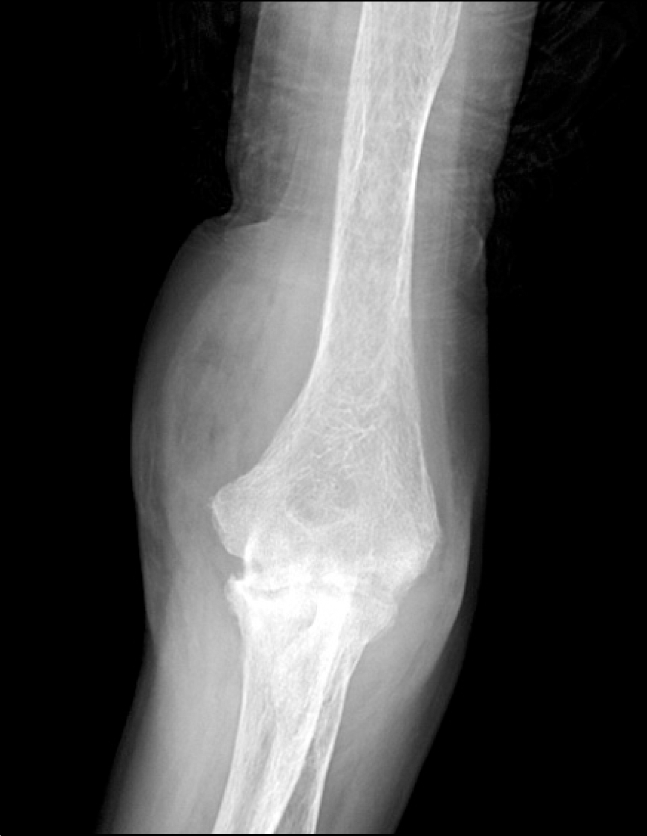
Figure 2.
Whole body bone scan of the patient who diagnosed as rheumatoid arthritis. Intense increased bone uptake in right wirst and left elbow, suggesting severe arthritis or septic arthritis.
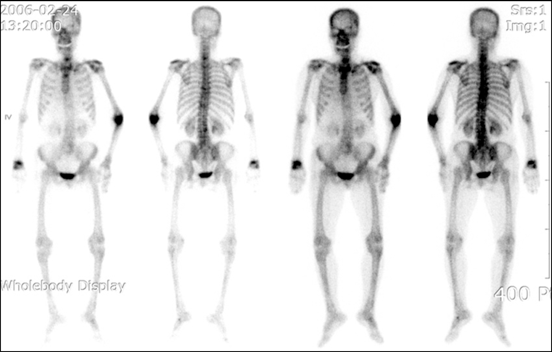
Figure 3.
Tuberculous arthritis of the Rt. 5th PIP joint. Radiograph demonstrated soft tissue swelling of 5 th finger. And enchondroma was shown at second proximal phalanx.
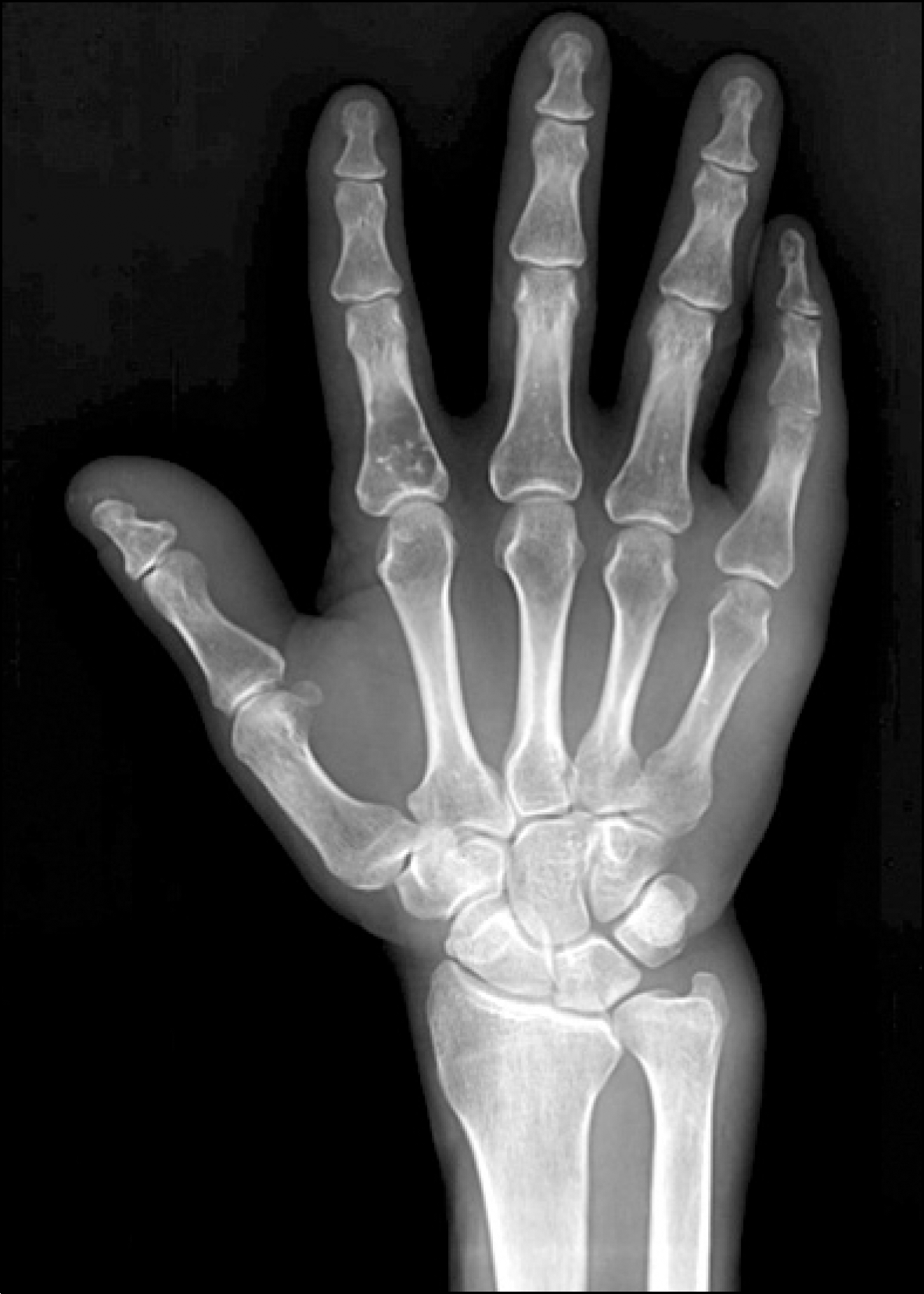
Table 1.
Baseline characteristics (N=39)
Table 2.
Characteristics of synovial pathologies
Table 3.
Comparisons between the diagnosed group and th undiagnosed group
| Diagnosed | Undiagnosed | p-value∗ | |
|---|---|---|---|
| Duration of synovitis (months, mean± SD) | 8.2±11.1 | 21.1±63.3 | 0.690 |
| ESR (mm/hr, mean± SD) | 26.5±34.4 | 10.4±14.3 | 0.419 |
| CRP (mg/L, mean± SD) | 24.7±22.8 | 6.5±8.9 | 0.207 |
| Techniques | |||
| Arthroscopic | 3 (8.6) | 32 (91.4) | 0.305 |
| Needle | 1 (25) | 3 (75) | |
| Mode | |||
| Spont | 4 (16) | 21 (84) | 0.287 |
| Trauma | 0 | 12 (100) | |
| Postop. | 0 | 2 (100) |
 XML Download