

 PDF
PDF ePub
ePub Citation
 Print
Print


Abstract
Backgrounds
Castration-induced androgen deprivation triggers a sequence of events, which activates apoptotic cell death of the androgen-dependent epithelial cells within the rat ventral prostate. To investigate the mechanism of castration-dependent apoptosis in the rat ventral prostate, the regulation of apoptosis-related genes was been investigated.
Methods
Azaline B was subcutaneously injected into Sprague-Dawley rat. The Fas receptor (Fas), Fas ligand (FasL) and bcl-2 mRNA, as well as the protein levels were detected by RT-PCR and Western blot analyses. Azaline B-dependent apoptosis was determined using TUNEL and a DNA fragmentation assay. The transacting factor of the FasL promoter was identified by DNA footprinting and a DNA mobility shift assay.
Results
The rat prostate was regressed after castration, with and the involuted ventral prostate regenerated by testosterone pretreatment, but not by that with FSH. Apoptosis of the ventral prostate was detected, after castration, using toluidine blue staining, a TUNEL assay and an apoptotic DNA fragmentation assay. The levels of Fas, FasL mRNA and protein were increased after castration. In the DNase I footprinting assay, using the FasL promoter and a nuclear extract prepared from a control prostate, at least two sites were protected: the SP-1 binding site at -283 bp and the prostate-unidentified factor (P-UF) binding site at -247 bp. The SP-1 binding activity vanished in the nuclear extract prepared from castrated rats. In the DNA mobility shift assay, the SP-1 binding activity was slightly decreased after castration. Both the Bcl-2 mRNA and Bcl-2 protein were downregulated after castration.
Figures and Tables
Fig. 1
Regression and regrowth of the rat ventral prostate gland in response to androgen manipulation. The ventral prostate obtained of 48 hours (A) or 5 days (B) after castration are shown, respectively The other assays were performed as described in Materials and Methods. Cast, castration; T, testosterone; FSH, follicle stimulating hormone.
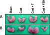
Fig. 2
Time course of the castration-dependent changes of prostatein C1 and TRPM-2 mRNA in the ventral prostate. At the indicated time, ventral prostate was removed from groups of rats which were either sham operated or castrated, and total RNA was prepared. Total RNA (30 µg) was analyzed by Northern blot hybridization using prostatein C1 and TRPM-2 cDNA probe as described in Materials and Methods.
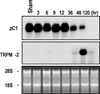
Fig. 3
Terminal deoxynucleotidyl transferase dependent dUTP nick end labeling (TUNEL) assay after castration. They were visualized by diaminobenzidine (DAB) and other cells were counter stained with methyl green as described in Materials and Methods. A, sham operation; B, castration; C, testosterone supplementation. Magnification: ×400.
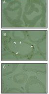
Fig. 4
Testosterone suppression of castration-induced apoptotic DNA fragmentation in rat ventral prostate. Before castration, testosterone (5 mg/day) were subcutaneously injected as 24 hrs intervals. After 48 hours, the rats were killed. a, DNA was isolated from ventral prostate, labeled on 3'-ends with [α-32P]-dideoxy-ATP, and analyzed by electrophoresis in 2% agarose gel. Autography and β-counting of low-molecular weight (≤15 kb) DNA fractions were followed. The amount of [α-32P]-dideoxy-ATP incoporated into DNA fractions from different groups were compared with that of intact animals at each time point an arbitrary unit 1.0. Cast, castration; Sham, sham operation; T, testosterone.
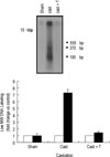
Fig. 5
Time course of castration-dependent change of FasL mRNA in rat ventral prostate. The FasL mRNA and protein levels were measured by RT-PCR and Western blot, respectively as described in Materials and Methods. PCR products were saperated on 1.2% agarose gel containing ethidium bromide. Animals were killed at indicated time after castration A, RT-PCR; B, Western blot.
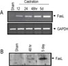
Fig. 6
Effect of testosterone and FSH on castration-induced FasL gene expression. Before castration, testosterone (5 mg/day) and FSH (20 IU/day) were subcutaneously injected as 24 hrs intervals. The FasL mRNA and protein levels were measured by RT-PCR as described in Materals and Methods. PCR products were separated on 1.2% agarose gel containing ethidium bromide. A, RT-PCR; B, Western blot. T, testosterone; AzB, azaline B.
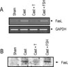
Fig. 7
Time course of castration-dependent change of Fas mRNA in rat ventral prostate. The Fas mRNA levels and protein were measured by RTPCR and Western blot, respectively, as described in Materials and Methods. PCR products were separated on 1.2% agarose gel containing ethidium bromide. Animals were killed at indicated time after castration A, RT-PCR; B, Western blot.
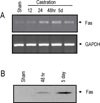
Fig. 8
Effect of testosterone and FSH on castration- induced Fas gene expression. Before castration, testosterone (5 mg/day) and FSH (20 IU/day) were subcutaneously injected at 24 hrs intervals. The Fas mRNA and protein levels were measured by RT-PCR and Western blot, respectively as described in Materials and Methods. PCR products were separated on 1.2% agarose gel containing ethidium bromide. A, RT-PCR; B, Western blot.
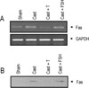
Fig. 9
The pattern of protein-binding site on the minimal FasL promoter region. The FasL promoter fragment (Hind III-Xho I) was labeled with [γ-32P]- dATP in the presence of T4 polynucleotide kinase at the -318 bp position of the coding, Upper strand. The labeled probe was incubated with 80 µg nuclear extracts from rat testis, and the mixtures were digested with 2 µL of DNase I (4,000 units/mL) for 30 minutes on the ice. After extraction with phenol/chloroform, the DNAs were resolved by electrophoresis on a 5% polyacrylamide gel in 8 M urea and the labeled DNA bands were visualized by autoradiography. The lane indicated by A+G is the wild type probe digested at adenine and guanine residues. The other assays were performed as described in Materials and Methods. A, DNase I footprinting; B, the sequences of 5'-flanking region of FasL gene. The arrow denoted the hypersensitive site. □, no protection; ■, protection.
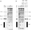
Fig. 10
Effect of castration on binding activity of nuclear extracts of prostate to SP-1 motif. Nuclear extracts from rat ventral prostate of sham and castrated were incubated with 32P-labeled double strand oligonucleotides corresponding to -293~-266 bp from the transcription initiation site. DNA-protein complexes were resolved native 4% polyacrylamide gel and visualized by autoradiography. The other assays were performed as described in Materials and Methods. Free did not contain nucear extracts. FD, Free DNA probe; Comp, competitor.
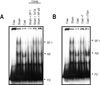
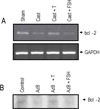
Fig. 11
Effect of testosterone and FSH on castration-induced Bcl-2 gene expression. Before castration and azaline B administration, testosterone (5 mg/day) and FSH (20 IU/day) were subcutaneously injected at 24 hrs intervals. The bcl-2 mRNA and protein levels were measured by RT-PCR and Western blot, respectively, as described in Materials and Methods. PCR products were separated on 1.2% agarose gel containing ethidium bromide. A, RT-PCR; B, Western blot.
References
1. Smith CA, Williams GT, Kingstone R, Jenkinson EJ, Owen JJT. Antibodies to CD3/T-cell receptor complex induce cell death by apoptosis in immature T cells in thymic cultures. Nature. 1995. 374:811–814.
2. Kerr JFR, Searle J, Harmon BV, Bishop CJ. Potten CS, editor. Apoptosis. Perspectives on Mammalian cell Death. 1987. Oxford University Press;93–94.
3. Tsujimoto Y, Shimizu S, Eguchi Y, Kamiike W, Matsuda H. Bcl-2 and Bcl- XL block apoptosis as well as necrosis: possible involvement of common mediators in aopototic and necrotic signa transduction pathway. Leukemia. 1997. 11:380–382.
4. Oehm A, Behrmann I, Falk W, Pawlita M, Maier G, Klas C, Li Weber M, Richards S, Dhein J, Trauth BC, Pansting IK, Krammer PH. Purification and molecular cloning of the APO-1 cell surface antigen, a member of the tumor necrosis factor/nerve growth factor receptor superfamily. Sequence identity with the Fas antigen. J Biol Chem. 1992. 267:10709–10715.
5. Suda T, Takahashi T, Golstein P, Nagata S. Molecular cloning and expression of the Fas ligand, a novel member of the tumor necrosis factor family. Cell. 1993. 75:1169–1178.
6. Nagata S, Golstein P. The Fas Death Factor. Science. 1995. 267:1449–1456.
7. Singer GG, Abbas AK. The Fas antigen is involved in peripheral but not thymic deletion of T lymphocytes in T cell receptor transgenic mice. Immunity. 1994. 1:365–371.
8. Tanaka M, Itai T, Adachi M, Nagata S. Down regulation of Fas ligand by shedding. Nat Med. 1998. 4:31–36.
9. Tanaka M, Suda T, Haze K, Nakamura N, Sato K, Kimura F, Motoyoshi K, Mizuki M, Tagawa S, Ohga S, Hatake K, Drummond AH, Nagata S. Fas ligand in human serum. Nat Med. 1996. 2:317–322.
10. Tanaka M, Suda T, Takahashi T, Nagata S. Expression of the functional soluble form of human Fas ligand in activated lymphocytes. EMBO J. 1995. 14:1129–1135.
11. Laouar Y, Sarukhan A, Pasqualetto V, Garcia C, Ezine S. Involvement of the Fas (CD95) system in peripheral cell death and lympoid organ development. Eur J Immunol. 1998. 28:1078–1088.
12. Shiraki K, Jsuji N, Shioda T, Isselbacher KJ, Takahashi H. Expression of Fas ligand in liver metastases of human colonic adenocarcinomas. Proc Natl Acad Sci USA. 1997. 94:6420–6425.
13. Suzuki A, Matsuzawa A, Iguchi T. Down regulation of Bcl-2 is the first step on Fas-mediated apoptosis of male reproductive tract. Oncogene. 1996. 13:31–37.
14. Isaacs JT. Antagonistic effect of androgens on prostatic cell death. Prostate. 1984. 5:545–557.
15. Chung WLK, McFadden DK. Sex steroid imprinting and prostate growth. Invest Urol. 1980. 17:337–342.
16. English HF, Kypianou N, Isaacs JT. Relationship between DNA fragmentation and apoptosis in the programmed cell death in the rat prostate following castration. The Prostate. 1989. 15:233–250.
17. Kyprianou N, English HF, Isaacs JT. Activation of a Ca2+-Mg2+-dependent endonuclease as an early event in castration induced prostatic cell death. Prostate. 1988. 13:103–117.
18. Lim K, Park C, Kim YK, Yun KA, Son MY, Lee YC, Park JI, Lee JH, Sul CK, Lee CS, Park SK, Hwang BD. Association of castration-dependent early induction of c-myc expression with a cell proliferation of the ventral prostate gland in rat. Exp Mol Med. 2000. 32:216–221.
19. Nickerson T, Pollak M, Huynh H. Castration- induced apoptosis in the rat ventral prostate is associated with increased expression of genes encoding insulin-like growth factor binding proteins 2, 3, 4 and 5. Endocrinology. 1998. 139:807–810.
20. Suzuki A, Masao E, Eguchi Y, Matsuzawa A, Nagata S, Tsujimoto Y, Iguchi T. Involvement of Fas in regression of vaginal epithelia after ovariectomy and during an estrous cycle. EMBO J. 1996. 15:211–215.
21. Taille de la A, Chen MW, Shabsign A, Bagiella E, Kiss A, Buttyan R. Fas antigen/CD-95 upregulation and activation during castration- induced regression of the rat ventral prostate gland. The Prostate. 1999. 40:89–96.
22. Tilly JL, Hsueh AJM. Microscale autoradiographic method for qualitative and quantitative analysis of apoptotic DNA fragmentation. J Cell Physiol. 1993. 154:519–526.
23. Lee J, Richburg JH, Younkin SC, Boekelheide K. The Fas system is a key regulator of germ cell apoptosis in the testis. Endocrinology. 1997. 138:2081–2088.
25. Virca GD, Northemann W, Shiels BR, Widera G, Broome S. Simplified northern blot hybridization using 5% sodium dodecyl sulfate. BioTechniques. 1990. 8:370–371.
26. Lim K, Yoo JH, Kim KY, Kweon GR, Kwak ST, Hwang BD. Testosterone regulation of proto-oncogene c-myc expression in primary Sertoli cell cultures from prepubertal rats. J Androl. 1994. 15:543–550.
27. Gorski K, Carneiro M, Schibler U. Tissue-specific in vitro transcription from the mouse albumin promoter. Cell. 1986. 47:767–776.
28. Lim K, Chae CB. Presence of receptor protein for testis-specific H2B (TH2B) histone gene in early stages of spermatogenesis. J Biol Chem. 1992. 267:15271–15273.
29. Kalb VF Jr, Bernlohr RW. A new spectrophtomeric assay for protein in cell extracts. Anal Biochem. 1977. 82:362–371.
30. Parker MG, White R, Williams JG. Cloning and characterization of androgen- dependent mRNA from rat ventral prostate. J Biol Chem. 1980. 255:6996–7001.
31. Montpetit ML, Lawless KR, Tenniswood M. Androgen repressed messages in the rat ventral prostate. Prostate. 1986. 8:25–36.
32. Schulze-osthoff K, Ferrari D, Los M, Wesselborg S, Marcus EP. Apoptosis signaling by death receptors. Eur J Biochem. 1998. 254:439–459.
33. Holz-Heppelmann CJ, Algreciras A, Badley AD, Paya CV. Transcriptional regulation of the human FasL promoter-enhancer region. J Biol Chem. 1998. 273:4416–4423.
34. McClure RF, Heppelmann CJ, Paya CV. Constructive Fas ligand gene transcription in Sertoli cells is regulated by Sp1. J Biol Chem. 1999. 274:7756–7762.
 XML Download