

 PDF
PDF ePub
ePub Citation
 Print
Print


Abstract
Background
Korean type 2 diabetic patients are known to differ from western diabetes because of their unique characteristics, such as non-obese but centrally obese anthropometry and relatively more insulin secretory defects than insulin resistance compared to western diabetic patients.
Methods
We recruited 1,646 diabetic patients in the present study and excluded the 45 patients with fasting C-peptide < 0.20 nmol/L. We had assessed insulin secretion by fasting serum C-peptide level and insulin resistance by short insulin tolerance test (Kitt; rate constant for plasma glucose disappearance, %/min) in the private diabetes clinic. The insulin secretory defect was divided by severe (C-peptide < 0.37 nmol/L), moderate (C-peptide 0.37~0.56 nmol/L), and normal (C-peptide ≥ 0.57 nmol/L) group. The insulin resistance was divided by insulin resistant (IR) (Kitt < 2.5 %/min) and insulin sensitive (IS) (Kitt ≥ 2.5 %/min) group.
Results
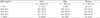
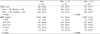
We analysed the data of 1,601 type 2 diabetic patients (831 men and 770 women, age 56.5 ± 10.8 years, duration of diabetes 9.6 ± 7.3 years). The prevalence of BMI ≥ 25.0 kg/m2 is 42.5% and BMI ≥ 23.0 kg/m2 is 70.2%. The prevalence of abdominal obesity (waist ≥ 90 cm in men and 80 cm in women) is 45.2% (36.0% and 55.2%, respectively in men and women). Fasting C-peptide level is 0.64 ± 0.29 nmol/L and Kitt value is 2.03 ± 0.96%/min. According to fasting C-peptide level, the degree of insulin secretory defect were severe (13.1%), moderate (33.0%) and normal (53.9%). According to Kitt value, the IR group is 70.6% and the IS group is 29.4%.
Figures and Tables


References
1. Min HK, Yoo HJ, Lee HK, Kim EJ. Changing patterns of the prevalence of diabetes mellitus in Korea. J Korean Diabetes Assoc. 1981. 6:1–4.
5. Zimmet P, Dowse G, Finch C, Serjeantson S, King H. The epidemiology and natural history of NIDDM-lessons from the South Pacific. Dia Metab Rev. 1990. 6:91–124.
6. Kopelman PG. Obesity as a medical problem. Nature. 2000. 404:635–643.
7. Flegal KM, Carroll MD, Ogden CL, Johnson CL. Prevalence and trends in obesity among US adults, 1999-2000. JAMA. 2002. 288:1723–1727.
13. Western Pacific Regional Office of the World Health Organization. The International Obesity Task Force. The Asia-Pacific perspective: redefining obesity and its treatment. 2000. In : Health Communications Australia; Sydney. http://www.obesityasiapacific.com.
14. Kim DJ, Lee MS, Kim KW, Lee MK. Insulin secretory dysfunction and insulin resistance in the pathogenesis of Korean type 2 diabetes mellitus. Metabolism. 2001. 50:590–593.
16. Lee TH. Prevalence of obesity in Korean non-insulin-dependent diabetic patients. Diabetes Res Clin Pract. 1996. 32:71–80.
 XML Download