

 PDF
PDF ePub
ePub Citation
 Print
Print


INTRODUCTION
Immunoglobulin E (IgE) is the lowest of 5 isotypes of human immunoglobulins (IgG, IgA, IgM, IgD, and IgE) in blood and has the shortest half life among them [12]. It primarily provides protective immunity against helminth parasites but can also respond to foreign substances even in small amounts and is accepted as a “gate keeper.” Under some conditions, IgE reaches pathological levels. Diseases which cause the elevation of serum IgE levels include atopic diseases (asthma, allergic rhinitis, atopic dermatitis, urticaria), parasitic diseases, cutaneous diseases, neoplastic diseases, and immune deficiencies [3]. Conditions associated with unusually high serum IgE concentrations (>1,000 IU/mL) are allergic bronchopulmonary aspergillosis, allergic fungal sinusitis, atopic dermatitis, human immunodeficiency virus infection, hyper IgE syndrome, IgE myeloma, lymphoma, systemic parasitosis and tuberculosis [4].
Some epidemiological studies showed that IgE levels were higher in subjects with cardiovascular diseases, in particular, in those experiencing unstable angina and acute coronary events [5]. But it is as yet unclear if the increased IgE level is a marker of future coronary incidents and whether it may be regarded as a risk factor of ischemic heart disease. A recent molecular study by Wang et al. [6] brought greater clarity to the relationship between high IgE levels and atherosclerosis showing that IgE plays an important role in the pathogenesis of atherosclerosis. However, clinical proofs demonstrating that high IgE levels could be a risk factor for an atherosclerotic outcome are still needed.
Our aim was to investigate the relationship between high serum IgE levels and some early atherosclerotic markers in patients without known atherosclerotic disease and risk factors.
MATERIALS AND METHODS
Study groups
The patients were randomly selected among outpatients referred to our allergy clinic with different symptoms and found to have high (>100 IU/mL) or very high serum IgE levels (≥1,000 IU/mL). Fifty patients (mean age, 40.96 ± 10.8 years; 22 female) who did not display any evidence of atherosclerotic disease and 30 healthy control subjects (mean age, 47 ± 8.27 years; 11 female) were included in the study. Allergic rhinitis, venom and food allergies were diagnosed with clinical symptoms, skin prick tests, and allergen specific IgE levels. Skin prick tests were done for Dermatophagoides farinea, Dermatophagoides pteronyssinus, grass pollen, weed pollen, tree pollen, Pariteria judaica, Artemisia vulgaris, Aspergillus furnigatus, Alternaria alternata (ALK Abello, Denmark). Allergen specific IgE levels for D. farinea, D. pteronyssinus, grass pollen, weed pollen, tree pollen, Pariteria judaica were measured by ImmunoCAP (Thermo Fisher Scientific Inc., Phadia AB, Uppsala, Sweden).
Skin tests were considered positive if there was a wheal response with a mean diameter of 3 mm or greater [7]. For immunoCAP a positive result was defined as value ≥0.35 kU/L [8].
Drug allergy was diagnosed with skin tests and oral challenge tests with commercial preparations according to the European Network for Drug Allergy recommendations [9].
Exclusion criteria
Patients with a previous history of arterial hypertension, diabetes mellitus, dyslipidemia, smoking, a diagnosis of ischemic heart disease and asthma were excluded from the study.
Measurement of total IgE, adhesion molecules, proinflammatory cytokines and hsCRP
Blood samples were obtained by puncturing the cubital vein. The serum was separated by centrifugation and stored at -80℃. Serum concentrations of total IgE (Siemens, Munich, Germany), intercellular adhesion molecule-1 (ICAM-1; Diaclone SAS, Besancon Cedex, France), vascular cell adhesion molecule-1 (VCAM-1; Diaclone SAS, Besancon Cedex, France), IL-6 (Diaclone SAS, Besancon Cedex, France), endothelin-1 (Biomedica Medizinprodukte GmbH & Co KG, Wien, Austria), and hsCRP (DRG international Inc., Springfield Township, NJ, USA) were determined by enzyme-linked immunosorbent assay.
Coronary flow measurements
Coronary flow reserve (CFR) recordings were performed with a Vivid 7 echocardiography device (General Electrics, Horten, Norway) using a middle-range frequency (3 to 8 MHz) broadband transducer. CFR recordings were performed in the left anterior descending coronary artery (LAD) by transthoracic Doppler echocardiography. The acoustic window was placed near the midclavicular line in the fourth and fifth intercostal spaces in the left lateral decubitus position. The left ventricle was imaged in the long-axis cross-section, and the ultrasound beam was inclined laterally. The coronary blood in the mid to distal LAD was identified by color Doppler flow mapping guidance with the optimal velocity range (+12 to +15 cm/sec). Then, the sample volume (1.5 or 2.0 mm wide) was positioned on the color signal in the LAD. Variables of LAD velocity were measured using fast Fourier transformation analysis. After baseline recordings of flow, dipyridamole (0.56 mg/kg) was infused over a 4-min period. An additional infusion of dipyridamole (0.28 mg/kg over a 2-min period) was used when the heart rate did not exceed the baseline by 10%. Two minutes after the end of the infusion, hyperemic spectral profiles in the LAD were recorded. All images were recorded for playback analysis and were later measured off-line. Average peak diastolic velocity (APDV) and average mean diastolic velocity were measured at the baseline and under hyperemic conditions. CFR was defined as the ratio of APDV at hyperemia to APDV at the baseline.
Carotid intima-media thickness measurements
The carotid arteries were evaluated with the Vivid 7 echocardiography device using a 10-MHz linear probe. The acquired images were recorded for playback analysis and were later measured offline. The common carotid artery, the carotid bulb, and internal and external carotid arteries were visualized on both sides. The intima-media thickness (IMT) of the carotid arteries was measured in the distal common carotid artery at a level 15 to 20 mm proximal to the carotid bulb. The 2 bright echogenic lines in the arterial wall were identified as the intima and the media. Three measurements were made for each side of the body; separate means were calculated and recorded as the right and left IMT.
Statistical analyses
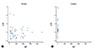
Data are presented as mean ± standard error. The statistical significance of the data was analyzed using SPSS ver. 17 (SPSS Inc., Chicago, IL, USA). The Kolmogorov-Smirnov test was used to evaluate the normal distribution of data. The differences between patients and control subjects were analyzed statistically using an independent sample t-test. CFR values of the patient and control groups were shown with a scatter distribution method. Statistical significance was acceptable to a level of p ≤ 0.05.
The study was approved by the ethical committee of Istanbul Faculty of Medicine and informed consent was obtained from all participants (approval number: 29449).
RESULTS
Patient characteristics
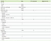
Thirty-four patients had an atopic disease while atopy was not found in the remaining 16 patients. The distribution of atopic diseases was as follows; allergic rhinitis (n = 16), food allergy (n = 7), drug allergy (n = 5), allergic rhinitis and concomittant food allergy (n = 4), venom allergy (n = 2) (Table 1).
Serologic investigation
Total IgE levels fell within a wide range in the patient group (range, 117–7,200 IU/mL; median, 8,610 IU/mL) while it was in close limits in the control group (range, 5.29–100 IU/mL; median, 29.7 IU/mL). The average total IgE level of atopic patients was 1,209 ± 1,031 IU/mL while it was 828 ± 757 IU/mL for nonatopics, which indicates no statistically significant difference (p = 0.194) (Table 2). There were no differences between the patient and control groups in terms of VCAM-1, ICAM-1, endothelin-1, and IL-6 levels, although hsCRP was higher in control subjects (Table 2).
CFR and carotid IMT measurements
CFR was significantly lower in the patient group when compared with the control group (p < 0.001; 95% confidence interval, -0.79 to -0.20) while carotid media thicknesses were not different among the 2 groups (Fig. 1).
There was no significant correlation between the CFR values and the different levels of total IgE, hsCRP, and other inflammation markers.
DISCUSSION
In this study, the CFRs of patients with no known atherosclerotic disease, but with high IgE levels due to atopic or unknown causes were found significantly lower when compared to those with normal IgE levels. This finding supports the hypothesis regarding the association between IgE and atherosclerosis.
Assessment of CFR constitutes a reliable and reproducible marker of coronary microvascular function however carotid IMT serves as a general measure of the degree of severity of atherosclerosis [1011]. Data demonstrating a relationship between coronary artery stenosis and CFR has grown [1213]. According to these studies, CFR has been considered as a sensitive marker for the identification of patients with high-risk anatomic disease by computed tomography angiography. Many studies demonstrated that even in patients without obstructive coronary artery disease (CAD), CFR is reduced for those with many conditions like hypertension, diabetes, metabolic syndrome, smoking, dyslipidemia, chronic kidney disease, and systemic inflammation when compared with healthy volunteers (The average CFR in these studies is 3.8.) [14]. These studies, as well as others, suggest that coronary microvascular dysfunction precedes the flow-limiting atherosclerotic plaque burden of epicardial coronary arteries in patients with risk factors. CFR reduction (<2.0) associated with cumulative coronary risk factors was present in up to 50% of a cohort of patients with suspected CAD and no evidence of ischemia. Recent studies, including large retrospective studies, consistently demonstrate that CFR is a significant prognostic marker in patients with known and suspected CAD [1516]. Low-risk patients classified by preserved CFR (≥2.0) are associated with a low risk for cardiac death (0.2% per year). In contrast, high-risk patients stratified by very low CFR (CFR < 1.5) are considered to be at high risk of cardiac death (11% per year). IMT of the common carotid arteries is a general measure of the severity of atherosclerosis, and increased IMT is related to generalized atherosclerosis. A number of studies have demonstrated the association of carotid IMT and the status of coronary atherosclerosis [1718]. In patients without clinical atherosclerotic disease, cardiovascular risk factors were found to be associated with impaired flow mediated dilation, CFR and increased IMT [18]. However, IMT values of the patient group were not different from the control subjects in another study [19].
Criqui et al. [20] first investigated the relationship between total IgE and cardiovascular disease. They found that mean total serum IgE levels increased 1.2 fold in men with a previous history of acute myocardial infarction (AMI). Subsequent studies, including both large and small series, demonstrated that different subgroups of ischemic heart disease, especially in men, was consistently associated with high serum IgE levels [21]. Accordingly, the hypothesis that IgE may play a role in the development of coronary heart disease has been put forward. Also, high IgE levels as a possible biomarker for ischemic heart disease has been discussed. Whether an increased IgE level is the cause or the result of atherosclerotic heart disease is still a matter of debate, as there is data supporting both sides. Szczeklik et al. [22] assessed the concentration of total serum IgEs in 100 patients with a recent AMI. The mean total IgE values increased steadily after the ischemic episode, achieving a statistically significant variation on day 3 and peaking on day 7. The concentration of IgE declined on day 14 and returned to the initial level after 3 weeks. Korkmaz et al. [23] also assessed the serum IgE values in 156 patients with coronary heart disease. They reported that the total serum IgE levels were significantly higher in patients with AMI and unstable angina. Szczeklik et al. [24] investigated the level of total serum IgE in 386 AMI patients at the time of a coronary care unit admission and divided the patients into the presence (n = 55) or absence (n=331) of sudden cardiac arrest. They found that the serum levels of total IgE were significantly higher in the former group. Edston and van Hage-Hamsten [25] studied 29 cases of sudden death because of coronary artery thrombosis. They reported that these patients had nearly twice the concentration of serum total IgE and a significantly greater frequency of increased total serum IgE levels.
All of these studies suggest that IgE levels increase as an immune response in acute coronary events. However, other studies propose that IgE itself may be a risk factor for ischemic heart disease. Langer et al. [26] measured IgE levels in 621 subjects who were then followed for an average of 9 years. The levels of total serum IgEs were found to be significantly higher in men who had experienced coronary heart disease, and also in those who experienced nonfatal AMI. This trend was not confirmed in women, however, in whom the levels of total serum IgEs were found to be nonsignificantly different from men. In another study, Kovanen et al. [27] assessed the association between total serum IgE levels and coronary heart disease in dyslipidemic men participating in the Helsinki Heart Study by following them for 5 years. They showed that basal IgE levels were significantly higher in those who had experienced a fatal or nonfatal AMI. A recent molecular and clinical study evaluating the pathogenesis of atherosclerosis further clarified the matters related to the relationship between IgE and atherosclerosis [18]. The concentration of total serum IgE was found to be nearly 60% higher in patients diagnosed with CAD than in those without and it was even higher in those with more severe CAD such as AMI than in those with unstable and stable angina. In the molecular part of the study IgE and the FcεR1 subunit FcεR1α were shown to be present in human atherosclerotic lesions, especially in macrophage rich areas. In mice, the absence of FcεR1α reduced inflammation and apoptosis in atherosclerotic plaques and reduced the burden of disease. In cultured macrophages, the presence of TLR4 was required for FcεR1 activity. IgE stimulated the interaction between FcεR1 and TLR4 which caused the apoptosis of the macrophages [6]. Taking together the results of these studies which demonstrated the proatherogenic, prothrombotic and antifibrinolytic effects of IgE, high IgE levels may be a risk factor for ischemic heart diseases [28].
The data obtained from human and animal studies which showed increased IgE levels as a risk factor for coronary heart disease were limited by only polyclonal IgE. The relationship between allergen-specific IgE and ischemic heart disease has not yet been shown. In a large and prospective investigation, Linneberg et al. [29] followed 18,841 people receiving subcutaneous allergen-specific immunotherapy (SCIT) along with 428,484 subjects receiving conventional allergy treatment (i.e., nasal steroids or oral antihistamines) for 10 years. As compared with conventional allergy treatment, patients receiving SCIT had an overall lower risk of mortality and AMI. This study suggested that allergen immunotherapy, which is known to alter the disease course as well as the immune response, may also reduce the risk of ischemic heart disease.
In healthy subjects and atherosclerotic patients some protein markers of vascular inflammation have been investigated as a noninvasive indicator of atherosclerosis. The most studied inflammation biomarker in cardiovascular disease is hsCRP [30]. hsCRP has been proposed as a strong biochemical marker in addition to other traditional risk factors such as dyslipidemia, hypertension, obesity and smoking [31]. However, other studies have shown that hsCRP does not play a direct role in atherogenesis. For example, hsCRP measurements of women and men (over 55 years) in the Rotterdam Study were reported as not providing a contribution in addition to the traditional risk factors [32]. Surprisingly, in our study hsCRP was found higher in control subjects. This finding can be explained by the fact that the patients had been treated with various drugs including antihistamines, nasal steroids or montelukast before being referred to our clinic which might have reduced inflammation.
Some other inflammatory markers such as VCAM-1, ICAM-1, and IL-6 were found to be associated with atherosclerosis. Ridker et al. [33] indicated that the plasma concentration of soluble ICAM-1 is elevated many years before the first myocardial infarction. However, only a few prospective studies have evaluated whether soluble VCAM-1 is also a marker for increased cardiovascular risk [34].
In this current study, none of the inflammatory markers were found to be different among the patient and control subjects, although the CFR values suggested an atherosclerotic tendency in the patient group. An explanation for this finding can be that this investigation was performed in a very early stage of a possible future atherosclerosis.
In conclusion, the results of this study seem to support the opinion that high total IgE levels may be a risk factor for atherosclerosis development. Future studies should investigate whether interventions to diminish IgE, such as anti-IgE treatment, can prevent atherosclerosis.

 XML Download