

 PDF
PDF ePub
ePub Citation
 Print
Print


INTRODUCTION
Ibuprofen is a common over-the-counter antipyretic-analgesic nonsteroidal antiinflammatory drug whose principal effect is inhibition of cyclooxygenase (prostaglandin-synthase), thus impairing the final transformation of arachidonic acid to prostaglandins, prostacyclin, and thromboxanes. Hypersensitivity-syndrome associated with ibuprofen is a host-dependent idiosyncratic drug-reaction [1].
Stevens-Johnson Syndrome (SJS) and toxic epidermal necrolysis (TEN) are life-threatening, bullous cutaneous diseases considered as immune-mediated reactions to drugs characterized by epidermal necrosis, extensive detachment of the epidermis, erosions of mucous membranes and severe constitutional symptoms [23]. This case describes a rare documented rapid-onset hypersensitivity of Ibuprofen causing SJS-TEN in a Nepali male.
CASE REPORT
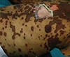
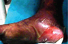
A 22-year-old Nepali male came to the Emergency Department of a teaching-hospital in central Nepal with chief complaint of sudden appearance of blisters on oral mucosa after taking Ibuprofen 400-mg tablets 8 hourly for 2 days for eye pain. He developed multiple maculae initially over trunk, then chest, face, lower limbs on the third day. He was admitted to the Emergency and upon examination, had extensive erythema (Fig. 1), necrosis, and exfoliating bullous detachment of the epidermis (Fig. 2) and mucous membranes (conjunctival, oral, and genital), with a positive Nikolsky's sign. The body temperature was 38℃, respiratory rate being 24 breaths/min, pulse rate 110 beats/min, blood pressure 130/90 mm Hg, SPO2 98%, and glucose random blood sugar 91 mg/dL.
The laboratory analyses are as follows: Na+, 138.8 mmol/L; K+, 4.02 mmol/L; urea, 41.0 mg/dL; creatinine, 1.0 mg/dL; platelet count, 60,000 mm3, white blood cell, 3,600×106/L; hemoglobin, 11.8 g/dL; prothombin (test), 17.5 seconds; prothombin (control), 13.0 seconds; international normalized ratio, 1.37; packed cell volume, 32.05%; bleeding time, 3 minutes; clotting time, 9 minutes; immunodeficiency virus I&II, nonreactive; hepatitis B surface antigen, nonreactive; urine routine examination, pus cells (8–10); C-reactive protein level, 340 mg/L; aspartate aminotransferase, 85 IU/L; alanine aminotransferase, 150 IU/L; total proteins concentration, 50 g/L; and albumin, 20 g/L.
Skin biopsy revealed prominent cell death with basal vacuolar changes and lymphocyte infiltrates with haziness in the dermo-epidermal junction which corroborated the clinical diagnosis of SJS-TEN.
Ibuprofen was immediately withdrawn and the patient was transferred to the intensive care unit (ICU). On admission to ICU, arterial blood gas analyses are as follows: pH, –7.442; pCO2, 28.5; pO2, 95.5; HCO3, 19.7; blood urea nitrogen, 17.0 mg/dL; blood-sugar random, 162.0; antinuclear antibody enzyme linked immune sorbant assay method, negative.
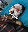
Patient had extensive sloughing off the face (Fig. 3) painful oral erosions with severe crusting of the lips and increased salivation. The plaques with vesicles, sloughed off more than 50% of body surface area which rendered difficulty to access vein on patients limbs. So, central venous pressure line insertion was done and enteral-nutrition was provided via a nasogastric tube. The patient's specific treatment with plasma exchange and fluid resuscitation with crystalloid infusions (1 mL/kg of body weight per hour) fine-tuned on the bases of the arterial blood pressure (>65 mm Hg), central venous pressure (≤10 mm Hg) and diuresis rate [4] was done. The basis for early plasma exchange was to remove/reduce the level of residual ibuprofen and its metabolites, critical cytokines (tumor necrosis factor-alpha [TNF-α], interferon-gamma [IFN-γ]), and drug-induced inflammatory mediators (perforin, granzyme-B released from cytotoxic T-lymphocytes and granulysin secreted by cytotoxic T-lymphocytes and natural killer cells) from the circulation [4]. However, the plasma exchange remain unsupported by controlled studies [5]. Subsequent systemic treatment included intravenous Ceftriaxone 1.0 gm 12 hourly (microbial findings on skin: Staphylococcus aureus and Pseudomonas aeruginosa; the symptoms and signs being warm, tender, erythematous, and edematous plaque over lower extremities with ill-defined borders), intravenous immunoglobulins (IVIG) 1.0-g/kg body weight/day infused over 6 hours for 3 successive days and corticosteroids 0.5-mg/kg body weight. Intravenous tramadol 25 mg was given for analgesia as per the patient's condition. The patient recovered completely after 5 weeks of intensive systemic and topical treatment.
DISCUSSION
SJS-TEN is a severe life threatening mucocutaneous syndrome caused by hypersensitivity to drugs and is associated with significant morbidity and mortality [6]. It is important to suspect the culprit drug by the temporal relationship (2 days to 8 weeks) [7] but the extremely 'rapid onset' in this case. The exact aetiopathogenesis being imprecise, is thought that some noxious metabolites, inflammatory mediators/modifiers, as well as cytotoxic T-lymphocytes, regulatory T cells and dermal-dendrocytes could provoke apoptosis/necrosis of epithelial cells [8910]. The human leucocyte antigen (HLA) system also plays an important role in the pathogenesis of TEN, since some drugs may bind directly to the HLA-complex and create self-reactivity due to the drug-modified HLA-peptide repertoire [10].
We must take in consideration that regional differences in drug prescription, the genetic background of patients (HLA, metabolizing enzymes) [1112], can have an impact on the incidence so a detailed drug history is extremely significant when go-getting to recognize the offender drug in SJS-TEN.
Initial symptoms of SJS-TEN can be unspecific like fever, stinging eyes and discomfort upon swallowing which precede cutaneous manifestations by a few days [13]. Early sites of cutaneous involvement are the presternal region of the trunk, face, palms, and soles. Erythema and erosions of the buccal, genital and/or ocular mucosa occurs in more than 90% of patients, with respiratory and gastrointestinal-tracts affected in few [14].
Prompt removal of causative drug should be the priority and the reported likelihood of a drug causing SJS-TEN can be found in PubMed/MEDLINE or the Litt's-drug-eruption reference manual [15]. Urgent plasma depuration (remain unsupported by controlled studies) [5] with supportive care by management of fluid and electrolyte requirements should be done. Wounds are treated conservatively, without skin debridement as blistered skin acts as a natural biological-dressing which favors re-epithelialization [13]. Topical and systemic medications (antibiotics if infection suspected, IVIG and corticosteroids) should be administered which prevents septicaemia and multisystem organ failure, the major cause of death in SJS-TEN. Recent case reports/series on the use of intravenous methylprednisolone with IVIG, and corticosteroids with infliximab and IVIG have been reported to be effective in arresting progression of TEN and reducing mortality [5].
IVIG, a highly purified IgG possessing anti-Fas activity, anti-infective properties also correcting protein and fluid loss, has for its support multiple, open-label, prospective studies, retrospective case series, and case reports as evidences [16].
 XML Download