

 PDF
PDF ePub
ePub Citation
 Print
Print


INTRODUCTION
Allergic bronchopulmonary aspergillosis (ABPA), a hypersensitivity respiratory disorder, is predominantly seen in asthmatics in the third and fourth decades of life. It is now well-recognised in India and is reported to occur in 7.6% of Indian adults with asthma [1]. Although ABPA is infrequently seen in children with asthma [2], it has been documented in infants as young as 6 months [3]. However, in children with cystic fibrosis, this entity is not as uncommon as in children with asthma [2].
The term "middle lobe syndrome" (MLS) was coined by Graham et al. [4] in 1948 to describe a clinical entity characterised by chronic or recurrent collapse of the right middle lobe while describing 12 patients with middle lobe atelectasis due to enlarged lymph nodes of nontuberculous origin. Brock et al. [5] originally described eight patients with recurrent atelectasis of the right middle lobe due to extrinsic compression by enlarged tuberculous lymph nodes and MLS caused by enlarged tuberculous lymph nodes is frequently known as "Brock's syndrome".
In ABPA, collapse, both lobar and segmental due to mucoid impaction is not infrequent but a MLS due to this disease is rather rare [678]. To our knowledge, ABPA as a cause of MLS has been documented in 3 adult patients [678] but never in children. We describe a 9-year-old female child with ABPA who presented with a MLS.
CASE REPORT
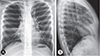
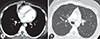
A 9-year-old female child was referred to our Institute for evaluation of right midzone patchy consolidation. She had episodic wheezing dyspnoea and nonproductive cough for 4 years, which had increased over the last fortnight with no acute distress, pallor, clubbing or cyanosis. Bilateral expiratory rhonchi were audible in all areas of the chest. The total leucocyte count was 7,190 cells/cumm, with neutrophils 68%, lymphocytes 25%, monocytes 2.6%, and eosinophils 4.4%. The absolute eosinophil count was 350 cells/cumm. In addition to the midzone patchy consolidation, an ill-defined opacity abutting the right cardiac border with loss of cardiac silhouette was detected on the chest radiograph (Fig. 1A). A right lateral view showed a wedge shaped density extending from the hilum anteriorly and inferiorly along with loss of volume confirming a MLS (Fig. 1B). High resolution computed tomography (HRCT) of the thorax confirmed MLS and also revealed central bronchiectasis (Fig. 2) which prompted investigations for ABPA. Skin prick test with antigens of Aspergillus fumigatus and Aspergillus flavus elicited a type I reaction. Strong bands of serum precipitins were detected against the same antigens. Specific IgE and IgG were positive for A. fumigatus and total serum IgE levels were elevated (1,488 IU/mL). Spirometry was suggestive of moderate obstruction with significant reversibility.
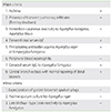
A diagnosis of ABPA presenting as MLS was made as she met 7/8 major diagnostic criteria (Table 1; Rosenberg-Patterson criteria [910]) and was initiated on oral prednisolone in the dosage of 0.5 mg/kg daily, which was tapered to alternate day dosages after a fortnight and tapered off after 6 months as the patient improved. She also received inhaled steroids for asthma. Marked symptomatic relief was noted within a fortnight with radiographic resolution of the MLS and reduction in total IgE levels to 903 IU/mL after 3 months. Informed written consent was obtained from mother.
DISCUSSION
MLS refers to chronic or recurrent collapse of the right middle lobe and has a distinct radiological presentation which continues to confound clinicians. Although, MLS is well documented in adults [11], this clinical entity is often overlooked in children with resultant diagnostic delay [12]. The occurrence of MLS in children with asthma is not infrequent and it is thought that this is caused by mucus hypersecretion which plug the bronchi [12].
It has been postulated that the propensity of the middle lobe to collapse may be due to the narrow diameter and long length of the middle lobe bronchus as well as the angular take-off of this bronchus. Furthermore, inadequate clearance of the impacted mucous along with the anatomically poor collateral ventilation contributes to the susceptibility of the middle lobe to collapse in isolation [1112].
The initial diagnosis of MLS is based on imaging and is distinctly recognised on a lateral radiograph as a wedge-shaped density extending from the hilum, anteriorly and inferiorly. However, it must be suspected on a posteroanterior radiograph, where the right cardiac border is obscured (Silhouette sign). A lordotic view would show the MLS as a wedge shaped density in the basal central zone of the right lower lung field, due to parenchymal involvement of the middle lobe. This view is often of help when there is diagnostic confusion. However, the HRCT can confirm the diagnosis as a trapezoidal or broad triangular opacity is visible with its base towards the hilum and is contiguous with the right cardiac border. In addition, it can detect other abnormalities like central bronchiectasis, as was seen in our patient [1112].
Although, ABPA is not an uncommon cause of lobar or segmental collapse, MLS has rarely been associated with this clinical entity [1]. A search of the literature revealed only three adult patients with ABPA who presented with MLS [678] but MLS as a presentation of childhood ABPA is yet to be documented. Our patient, a female child met 7/8 major diagnostic criteria for ABPA including central bronchiectasis, a pathognomic feature central to the diagnosis [1]. All 3 documented patients had marked symptomatic improvement after therapy with oral corticosteroids [678]. In 2 patients [67], there was radiological clearance but in the patient [8] documented by us in 2014, MLS persisted till the time of publication. However, a review at 9 months revealed an inflated middle lobe and radiological clearance. Our patient too had a marked symptomatic and radiological clearance after therapy with oral corticosteroids.
In an appropriate setting, all children with Aspergillus sensitive asthma should be evaluated for ABPA.
 XML Download