

 PDF
PDF ePub
ePub Citation
 Print
Print


INTRODUCTION
Hamano et al. [1] first reported the association of elevated serum immunoglobulin G4 (IgG4) levels in patients with autoimmune pancreatitis in 2001. A syndrome comprising, in addition to sclerosing pancreatitis, sclerosing sialadenitis and/or retroperitoneal fibrosis was subsequently described [2, 3]. Other clinical manifestations including autoimmune hepatitis [4], inflammatory pseudotumour [5], nonspecific interstitial pneumonia [6] and inflammatory abdominal aortic aneurysm [7] have also been linked with elevated serum levels of IgG4. It has, therefore, been suggested that the serum IgG4 concentration is useful in diagnosing autoimmune pancreatitis and Mikulicz disease [1, 8, 9, 10]. A serum IgG4 concentration greater than 1.3 g/L has been reported to confer a sensitivity of 66.7% to 94.3% and specificity of 93% to 97% for the diagnosis of autoimmune pancreatitis [1, 8, 9, 11]. In contrast, Ghazale et al. [11] showed that elevated IgG4 levels have a poor positive predictive value of 36% when 1.4 g/L was used as a cutoff. In other manifestations of IgG4-related sclerosing disease, the diagnostic utility of serum IgG4 concentrations is not known.
We have observed a 15% increase per annum in the number of requests for serum IgG4 and IgG subclass concentrations over the last several years. This may be due, at least in part, to a growing awareness of this new clinicopathological entity. Therefore, we sought to determine the utility of serum IgG4 concentration in the diagnosis of IgG4-related sclerosing disease by determining the clinical characteristics of unselected patients with elevated serum concentrations of IgG4. We also analysed other laboratory parameters to see if their inclusion improved diagnostic accuracy for the syndrome.
MATERIALS AND METHODS
The study was approved by the Human Research and Ethics Committee of the Sydney South West Area Health Service. Between January 2009 and June 2010, we performed 1,258 IgG subclass concentrations as a part of routine diagnostic service when either IgG4 levels alone or IgG subclass concentrations were requested. These were performed by nephelometry (Immage 800, Beckman Coulter Inc., Brea, CA, USA) using Human IgG Subclass reagents (The Binding Site Group Ltd., Birmingham, UK). The clinical diagnosis of all patients with elevated IgG4 concentrations, defined as >1.30 g/L, was then obtained from clinical records. Patients with suggestive histological findings and/or convincing clinical features, which included typical imaging and responses to steroids, were diagnosed with IgG4-related sclerosing disease. If there was insufficient clinical information to determine a likely diagnosis, the sample was excluded from the study. The serum IgG4 to total IgG ratio as well as eosinophil counts, serum IgE, IgA and IgM concentrations, C-reactive protein (CRP) and erythrocyte sedimentation rate (ESR) were analysed when available and correlated with the clinical diagnosis. Data were analysed using GraphPad Prism ver. 5.02 (GraphPad Software Inc., La Jolla, CA, USA); statistical analyses were performed using Mann-Whitney test; p < 0.05 was considered statistically significant. The confidence interval for the positive predictive value was determined by exact confidence intervals.
RESULTS
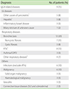
Of 1,258 IgG subclass concentrations performed, 80 patients (6.4%) had a IgG4 concentration greater than 1.30 g/L. Analysis of the records of 61 (40 males and 21 females) of these patients was performed as adequate clinical data were not available for 19 patients. Of the 61 patients, 9 patients (5 males and 4 females) had convincing clinical features of IgG4-related sclerosing disease and 5 of these had histological features consistent with the diagnosis. The median age of those with and without IgG4-related sclerosing disease was 58 and 52 years, respectively (p = 0.14). The manifestations of IgG4-related sclerosing disease included autoimmune pancreatitis (3), retroperitoneal fibrosis (3), sclerosing sialadenitis (1), lacrimal gland enlargement (1), orbital pseudotumour (1) and sclerosing cholangitis with lymphadenopathy (1). One patient with autoimmune pancreatitis also had retroperitoneal fibrosis. All of these patient were either first diagnosed with IgG4-related sclerosing disease based on biopsy results or their treating clinician had a high index of suspicion for the syndrome on clinical grounds prior to IgG4 testing. The characteristics of those who did not have IgG4-related sclerosing disease but had elevated IgG4 concentrations are described in Table 1.
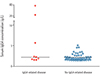
The median serum IgG4 concentrations of those with and without IgG4-related sclerosing disease were not significantly different at 2.16 g/L and 1.86 g/L respectively (p = 0.22) (Fig. 1). To determine if patients with IgG4-related sclerosing diseases had a disproportionate increase in that subclass relative to other subclasses, we analysed the serum IgG4 to total IgG ratio in each group. The median value of serum IgG4 to total IgG ratio in those with and without IgG4-related sclerosing disease was similar at 0.121 and 0.118 respectively (p = 0.09). There were also no significant differences between the eosinophil counts, CRP, ESR, serum IgE, IgA, and IgM concentrations of two groups where the information was available (data not shown).
The positive predictive value of serum IgG4 concentrations for the diagnosis of IgG4-related sclerosing disease was 15% (95% confidence interval, 7-26%). If a serum IgG4 concentration of 2.8 g/L was used as a cutoff, the positive predictive value still remained low at 23% (3/13). The sensitivity and specificity could not be determined as patients who had IgG4 concentrations ≤1.3 g/L were not clinically categorised.
DISCUSSION
High serum IgG4 concentrations are reported to have good sensitivity and specificity for the diagnosis of IgG4-related sclerosing disease but, because of the low prevalence of the condition, the positive predictive value of this test is reported as between 36-79%, depending on the sample populations [1, 8, 9, 11, 12]. These studies have used other pancreatic or salivary diseases other than those diagnosed with IgG4-related sclerosing disease as control populations and subsequently, the proportion of those with this syndrome is over-represented, giving a much higher positive predictive value.
The value of high serum IgG4 concentrations in an unselected population has recently been assessed in other similar studies [13, 14]. Consistent with these studies, our findings in a tertiary hospital cohort referred for measurement of IgG subclasses confirm that the yield for diagnosis of IgG4-related sclerosing disease is low at 15% with a cutoff of 1.3 g/L. In the study by Ghazale et al. [11], the positive predictive value could be improved to 75% if a serum IgG4 concentration of 2.8 g/L was used. However, in the cohort reported here, the positive predictive value still remained poor at 23% even after increasing the cutoff value to 2.8 g/L. Therefore, elevated serum IgG4 concentrations must be interpreted with caution for the diagnosis of IgG4-related sclerosing disease especially when the pretest probability of the disease is low and IgG4-immunostaining of an involved tissue is nondiagnostic or not available.
Previous studies have indicated that serum IgG4 to total IgG ratio was significantly elevated in IgG4-related sclerosing diseases [10, 15]. However, in the patients with elevated serum IgG4 concentrations reported here, we did not find this ratio to be more useful than absolute levels in differentiating those with and without IgG4-related sclerosing disease. The previously reported elevation of serum IgG4 to total IgG ratio in IgG4-related sclerosing disease is likely due to an increase in absolute IgG4 concentration that is not specific to this disease, hence adding no further diagnostic value.
In their study, Kanari et al. [16] reported that all nine patients with IgG4-related lacrimal gland enlargement had elevated serum IgE levels and the IgE levels significantly correlated with IgG4 concentrations. Five of 9 patients also had eosinophilia and asthma-like symptoms. These findings were explained by postulating that enhanced Th2 cell differentiation and interleukin (IL) 10 production is involved in the pathogenesis of IgG4-related lacrimal gland enlargement. However, in our cohort with high serum IgG4 concentrations, we did not find significantly higher IgE levels or eosinophil counts in those with IgG4-related sclerosing disease compared to those without the disease. It is likely that, while Th2 cell differentiation and IL-10 production is critical for IgG4 production, it also occurs in other situations where IgG4 production is increased.
In our cohort, a number of patients with elevated serum IgG4 concentrations without IgG4-related sclerosing disease had respiratory diseases, in particular, bronchiectasis (Table 1). This could be explained because the samples may have been referred to exclude immunodeficiency as an explanation for suppurative lung disease. In this context, it is interesting that Shakib et al. [17] showed that 7/16 patients with cystic fibrosis had elevated IgG4 concentrations. Whether a significant proportion of bronchiectatic patients also has high IgG4 concentrations has not been reported. The IgG4 subclass response is known to become prominent with chronic antigenic stimulation for certain antigens such as honey bee venom [18]. While this may not be the case for all antigens, such chronic stimulation would explain the observed elevated IgG4 concentrations in patients with chronic infectious states and other inflammatory conditions (Table 1).
It is not surprising that IgG4 levels have a low positive predictive value for the diagnosis of IgG4 related disease since this subclass is elevated in other conditions also. For example Lin and Li [19] reported elevations of IgG4 as part of the hypergammaglobulinaemia detected in both active and inactive rheumatoid arthritis and it is likely that other inflammatory conditions also will have elevations of all IgG isotypes. Similarly, our findings indicate that high levels of IgG4 can also be found in nonautoimmune pancreatitis (Table 1).
There were six patients in our cohort who had elevated IgG4 concentrations without apparent IgG4-related sclerosing disease but rather had autoimmune hepatitis, biliary stricture of unknown cause, unexplained nephritis or interstitial lung disease. These conditions have previously been described in association with elevated IgG4 concentrations [4, 6, 20, 21]. Thus, it is possible that we have underestimated the positive predictive value by excluding these patients. However, even if this was the case, the positive predictive value would still remain poor at 25%.
Our study is limited by its retrospective study design and by a relatively small number of individuals with criteria that fulfilled the definition of IgG4-related sclerosing disease. In addition we have a referral bias as 6.4% of our patients had IgG4 concentrations greater than 1.3 g/L compared to an expected proportion of 2.5% based on analysis of values for a healthy adult control population [22]. This can be explained by the fact that, as a tertiary hospital immunology laboratory, we tend to receive more samples from patients with various inflammatory conditions rather than healthy individuals. This referral bias can affect the positive predictive value in both directions; as some patients were referred for an assessment of other unrelated conditions such as immunodeficiency, this could lower the positive predictive value. On the other hand, a number of patients were referred specifically for measurement of IgG4 levels with the possibility of IgG4-related sclerosing disease in mind and thus, it is possible that the true positive predictive value in the unselected population is even lower. Despite these shortcomings the large proportion of patients with elevated IgG4 without evidence of the features of IgG4-related sclerosing disease is an important observation.
In conclusion, serum IgG4 concentration has a poor positive predictive value in the diagnosis of IgG4-related sclerosing disease. While serum IgG4 concentration still has valuable diagnostic utility when the pretest probability of the disease is high, indiscriminate testing of serum IgG4 levels should be discouraged as it does not add diagnostic value in the absence of consistent clinical findings. Furthermore, high serum IgG4 concentrations in the absence of histological staining should be used with caution in diagnosing IgG4-related sclerosing disease as a number of conditions with chronic antigen stimulation may also result in high IgG4 concentration. Finally, as the pathogenesis of IgG4-related sclerosing disease still remains unknown, care should be taken not to overemphasise the role of IgG4 in describing new disease associations.
 XML Download