

 PDF
PDF ePub
ePub Citation
 Print
Print


INTRODUCTION
Asthma is chronic inflammatory allergic disorder which affects numbers worldwide. The treatment strategy for asthma consists of allergen-avoidance, pharmacotherapy, patient education and immunotherapy [1]. Allergen immunotherapy, which was first developed by Noon and Freeman more than 100 years ago, has a unique role in the treatment of allergic diseases in that only immunotherapy can induce long-term immunological tolerance [2, 3]. Allergen immunotherapy is indicated in patients with IgE-mediated allergic diseases such as allergic rhinitis, allergic asthma, and insect venom anaphylaxis. Subcutaneous immunotherapy (SCIT) has been used as a majority in the field of clinical practice and research. Now sublingual immunotherapy (SLIT) which shows similar clinical effectiveness is also commercially available [4].
Successful outcomes with immunotherapy are not only reductions in symptoms, medication requirements and improved quality of life, but also long-term tolerance. Immunological changes such as induction of regulatory T cell (Treg), induction of a blocking antibody, IgG4, with reduction of IgE production (the shift in balance between IgE and IgG4), and decrease of tissue mast cell and eosinophil are well-known as the mechanism of immunotherapy [5]. Clinically, its early effects are reduction in symptoms and need for medication, which are followed by further reduction in symptoms and need for medication but also reduction in hyperresponsiveness and late-phase response [6]. Even after end of treatment period, immunotherapy provides persistent effects of long-term reduced symptoms/ need for medication as well as long-term reduced hyperresponsiveness and late phase response [6]. Immunotherapy also provides preventive effect: it seems not only to reduce the development of new allergic sensitivities as measured by skin prick test or allergen-specific IgE but also to provide preventive effect on later development of asthma in children with seasonal rhinoconjunctivitis [6, 7].
It would be useful to develop animal models to elucidate pathophysiologic mechanisms and to develop new treatment modalities. Mouse models have many advantages such as well-known immune system, availability of many tools to manipulate immunological process, numerous genetically modified (transgenic or knockout) strains, and economical aspects [8]. We have previously published immunotherapy models using CpG-oligodeoxynucleotides and oral desensitization in mice [9-12]. In this study, we tried to establish murine immunotherapy models which have similar findings in human using a subcutaneous rush immunotherapy-like protocol.
MATERIALS AND METHODS
Mice and asthma model
Six-week old female Balb/c mice were purchased from DBL (Korea). Balb/c mice were maintained in the clinical research institute at Seoul National University Hospital. All animal experiments were approved by the committee on animal experimentation at our institution.
Sixteen mice were used for the determination of the maximal tolerable dose and another eight mice in each group for the experiment of immunotherapy. Mice were sensitized by injection of 20 µg ovalbumin (OVA, Grade V; Sigma, USA) emulsified in 1 mg aluminum hydroxide intraperitoneally. Animals received an identical booster immunization 14 days later. On days 21, 22, and 23 after the initial sensitization, mice were challenged for 30 min with an aerosol of 1% (w/v) OVA in phosphate buffered saline (PBS) in a Plexiglas chamber using an ultrasonic nebulizer (NE-U12; Omron, Japan) as previously described [12].
Determination of the tolerable doses of OVA subcutaneous immunotherapy
One week af ter the las t challenge, mice were given subcutaneous injections of OVA from the dose of sensitization (20 µg) that was increased in two folds at 12 h interval to determine the tolerable dosage of OVA immunotherapy. The appearance, body weight, activity and behaviors were observed. Maximal tolerable dose was determined as the dose which did not cause harmful effect on the animal. Maximal safe dose was determined as the dose which did not cause any significant harmful effect on the animal.
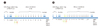
Schedule of OVA subcutaneous immunotherapy (short term, 10 times) and rechallenge
We used both of the maximal tolerable dose and the maximal safe dose for the experiment. Mice were given subcutaneous injections of OVA from the dose of sensitization (20 µg) that was increased in two folds at 12 h interval to reach the maximal tolerable dose or the maximal safe dose. After that, mice were injected with each dose according to the group for total of 10 times. Ten mice belonged to each group.
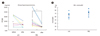
Mice were rechallenged with OVA inhalation for three days one week after the final injection of subcutaneous immunotherapy in the same way of the initial OVA challenge (Fig. 1A).
Schedule of OVA subcutaneous immunotherapy (prolonged, 24 times) and rechallenge
Mice were given subcutaneous injections of OVA from the dose of sensitization (20 µg) that was increased two-fold at 12 h interval to reach the maximal tolerable dose. After that, mice were injected with the maximum tolerable dose for total of 24 times. PBS was injected for the controls. Eight mice belonged to each group.
Mice were then rechallenged with OVA inhalation for three days one week after the final injection of subcutaneous immunotherapy (Fig. 1B).
Airway hyperresponsiveness (AHR)
Twenty-four hours after each final OVA inhalation challenge, AHR was assessed by determining methacholine-induced airflow obstruction (Penh) using one chamber whole body plethysmography (Allmedicus, Korea), as previously described [12]. Increasing doses of methacholine (ranging from 2.5-50 mg/mL; Sigma, USA) were administered by nebulization for 3 min, and Penh values were calculated over the subsequent 3 min. Results were presented as PC200 values, which are defined as the concentration of methacholine required to increase baseline Penh by 200%.
Bronchoalveolar lavage (BAL)
Forty-eight hours after each third OVA challenge, tracheae were cannulated and lungs were lavaged with five 0.4 mL aliquots of pyrogen-free saline. After Diff-quickR staining lung lavage cells in cytospin preparations, two investigators counted blindly more than 300 inflammatory cells under a light microscope [12].
Antibody responses
Forty-eight hours after each third OVA challenge, blood samples were obtained by cardiac puncture. Antibody titers were measured as previously described [12]. Briefly, microtiter plates (Dynex Technologies, USA) were coated overnight with 2 µg/mL of OVA in a 50 mM carbonate buffer (pH 9.6) at 4℃. Nonspecific binding was blocked with 2% bovine serum albumin for 1 h at 20℃. After incubating with test sera for 2 h, plates were incubated with horse radish peroxidase-labeled goat anti-mouse IgE or IgG2a (PharMingen, USA) for 1 h at 20℃. The reaction was developed using a tetramethylbenzidine (Sigma, USA) substrate and then stopped by adding 2 N H2SO4. Subsequently, optical density was measured at 450 nm. A high titer of anti-OVA IgE or IgG2a was used as a standard, and linear standard curves were obtained by serially diluting standard serum. The results are expressed in arbitrary units according to measured OD values.
Cytokine production by splenocytes
Cytokine production by splenocytes was evaluated as previously described [9]. Briefly, spleens were homogenized using a 94-µm screen (Bellco Glass Inc., USA) to obtain single cell suspensions. Splenocytes (2×10⁶) were then cultured with OVA (100 µg/mL) or PBS control in 12-well plates. After 2 days, IL-4, IL-5, and INF-γ production levels were quantified in culture supernatants by sandwich ELISA using specific monoclonal antibody pairs as the manufacturer's guide.
RESULTS
Determination of the doses of OVA subcutaneous immunotherapy
One week af ter the initial challenge, mice were given subcutaneous injections of OVA from the dose of sensitization (20 µg) that was increased in two folds at 12 h intervals to determine the tolerable dosage of OVA immunotherapy. The appearance, body weight, activity and behaviors were monitored. Maximal tolerable dose was determined as the dose which did not cause any significant harmful effect on the animal.
Eight-fold increase (160 µg) did not affect any feature of mice with normal activity. Sixteen-fold increase (320 µg) did not affect major significant harmful effect but caused slightly reduced activity in half of the group (8/16). Thirty two-fold increase caused reduced activity. The doses of ×64 to ×128 caused reduced activity and dyspnea. The dose of ×250 caused mortality in more than half of the group (5/8) within 30 min after OVA injection. The maximal tolerable dose was determined as sixteen-fold increase (320 µg) and maximal safe dose was determined as eight-fold increase (160 µg) from the sensitization dose.
Effects of OVA subcutaneous immunotherapy (short term, 10 times)
Antibody responses
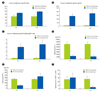
There was no significant difference in serum OVA specific-IgG1 level between before and after the short-term immunotherapy with either doses of eight-fold increase (160 µg) or sixteen-fold increase (320 µg). However, there was significantly increased serum OVA specific-IgG2a production after the short-term immunotherapy with both of the doses (Fig. 2). The balance of Th1 and Th2 response was further evaluated with the parameter of IgG2a/IgG1 ratio as previously described [13]. The IgG2a/IgG1 ratio was significantly increased after the short-term immunotherapy with both of the doses, which meant that the balance was towards Th1 immune response (Fig. 2).
Cytokine production by splenocytes
IL-5 production from splenocytes was significantly decreased after the short-term immunotherapy with either doses of eight-fold increase (160 µg) or sixteen-fold increase (320 µg) (Fig. 2). IL-4 was unfortunately not detected in this experiment.
INF-γ production from splenocytes was significantly decreased after the short-term immunotherapy with dose of eight-fold increase (160 µg) while there was no difference after the short-term immunotherapy with dose of sixteen-fold increase (320 µg). So, the balance of Th1 and Th2 response was further evaluated with a parameter of IL-5/INF-γ ratio. Interestingly, IL-5/INF-γ ratio was decreased after the short-term immunotherapy with dose of sixteen-fold increase (320 µg) while there was no difference after the short-term immunotherapy with dose of eight-fold increase (160 µg), which was different finding compared with that in antibody responses (Fig. 2).
AHR
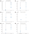
There was no significant difference in AHR between before and after the short-term immunotherapy with either doses of eight-fold increase (160 µg) or sixteen-fold increase (320 µg) (Fig. 3A).
BAL fluid and airway inflammation
There was no significant difference in airway inflammatory cell profiles including eosinophils in BAL fluid before and after the short-term immunotherapy with either doses of eight-fold increase (160 µg) or sixteen-fold increase (320 µg) (Fig. 3B).
Effects of OVA subcutaneous immunotherapy (prolonged, 24 times)
Antibody responses
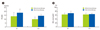
There was a significant decrease in serum OVA specific-IgE and IgG1 level after the prolonged immunotherapy with high dose (16-fold higher than the dose of sensitization) OVA (320 µg). Interestingly, there was also significantly decreased serum OVA specific-IgG2a production after the prolonged immunotherapy as well (Fig. 4).
Cytokine production by splenocytes
IL-5 production from splenocytes was significantly decreased after the prolonged immunotherapy. IL-4 seemed also to be decreased but not significantly.
INF-γ production from splenocytes was significantly decreased after the prolonged immunotherapy (Fig. 4).
AHR
Prolonged immunotherapy showed a protective effect on AHR; while AHR was increased in PBS treated group after OVA rechallenge, there was no significant difference in AHR in immunotherapy group (Fig. 5A).
BAL fluid and airway inflammation
There was no significant difference in airway inflammatory cell profiles including eosinophils in BAL fluid after the prolonged immunotherapy (Fig. 5B).
DISCUSSION
Short term immunotherapy (10 times) with OVA at the maximal safe and tolerable dose decreased IL-5 production, decreased IL-5/INF-γ ratio, and increased IgG2a/IgG1 ratio but there was no significant difference in AHR or airway inflammation. Prolonged immunotherapy (24 times) with at the maximal tolerable dose not only decreased cytokine productions of IL-5 and even INF-γ, but also decreased OVA-specific IgE, IgG1 and even IgG2a productions. Remarkably, worsening of the AHR after rechallenge was inhibited after the prolonged immunotherapy.
Immunotherapy in allergic disease, which was introduced 100 years ago, is an effective treatment modality in IgE-mediated diseases such as asthma, allergic rhinitis and venom allergy. Immunotherapy is the only treatment modality with the potential for long-term immunologic amelioration of allergic diseases [14]. However, a number of important questions such as units of allergen potency, treatment schedule, identification of patients group who show effectiveness, safety, long-term effects, possible new indications, and developments in the production of allergens remain unanswered [15].
Mechanism of allergen immunotherapy is suggested as follows [5]. Initially decrease in mast cell and basophil degranulation soon after immunotherapy followed by generation of allergen-specific Treg cells and suppression of effector cells. In terms of antibody reactions, allergen-specific IgE levels show an early increase and a late decrease. A blocking antibody, allergen-specific IgG4, increases from relatively early phase. Type 1 skin test reactivity will be decreased later. The numbers of tissue mast cell and eosinophil will decrease after a few months [5].
As we have previously published immunotherapy models using CpG-oligodeoxynucleotides and oral desensitization [9-12], we tried to establish immunotherapy models with allergen only using subcutaneous rush immunotherapy-like protocols. In this study, we established rush immunotherapy-like schedule with maximal tolerable dose and showed that short term immunotherapy model is comparable to the initial response of allergen immunotherapy in human; early induction of blocking antibody (IgG2a in this study) instead of decreased IgE response and suppression of Th2 cytokine, IL-5. Especially, this study showed that IgG2a/IgG1 or IL-5/INF-γ ratio improved dramatically from early phase with repeated subcutaneous OVA injections. The decrease in IgE/IgG4 ratio during SCIT seems to be a feature of skewing from allergen-specific Th2 to Treg cell predominance in human since the class switching of IgG4 is caused by the co-stimulation of IL-4 and IL-10 [5]. IgG1/IgG2a or IL-5/INF-γ ratio could be good markers indicating early immunological changes reflecting the shift of Th2/Th1 balance during immunotherapy in mice.
It was interesting that OVA-specific IgG2a and INF-γ productions as well as OVA-specific IgE/IgG1 and IL-5 production were also decreased after prolonged immunotherapy. Prolonged immunotherapy seems inhibit both Th2 and Th1 responses to OVA. Prolonged immunotherapy also provided some physiological benefit of inhibiting the enhancement of AHR.
Inhibition of both Th1 and Th2 responses by high dose subcutaneous immunotherapy was a new finding. The murine models of immunotherapy in this study could be helpful to develop immunotherapy protocols and to evaluate the mechanism of the induction of tolerance by immunotherapy. We could not develop the protocols which could reverse or treat whole figures of asthma including eosinophilic airway inflammation and AHR. More prolonged immunotherapy may be needed to see these effects. We did not evaluate the regulatory cytokines such as IL-10 or TGF-β in this study which would be the limitations of this study. These cytokines are very critical to understand the underlying mechanisms of immunotherapy. However, we described here proposed models of SCIT in murine asthma model, which showed some beneficial immunological and physiological changes. Further studies are needed to elucidate the underlying mechanism of immunotherapy, which would provide more improved knowledge of the induction of tolerance by the immunotherapy.
 XML Download