

 PDF
PDF ePub
ePub Citation
 Print
Print


INTRODUCTION
Autoerythrocyte sensitization syndrome (AES) was first described by Gardner and Diamond in 1955 [1], when four women with painful bruising were depicted. Patients with AES typically present with the development of recurrent, spontaneous, painful ecchymosis, frequently preceded by a prodrome of pain or itching of the skin. Over 100 cases have been reported since then [2], and nearly all cases were in women [3, 4]. The patients are sensitive to their own red blood cells injected intradermally, and underlying coagulopathies are thought to be absent. Psychophysiologic complaints are so frequent that AES has also been referred to as psychogenic purpura.
CASE REPORT
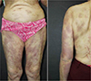
A 70-year-old woman had recurrent episodes of painful bruising on the back, abdomen, and four extremities for 5 years (Fig. 1). The patient complained that the bruises appeared after scratching the skin to relieve pruritus. The patient had a 5-year history of general chronic neurodermatitis without any bleeding manifestations, such as melena, hematuria, and menorrhagia. The neurodermatitis had usually recurred approximately every 4-6 months with severe itching over the lesions, and the last relapse was approximately 3 months prior to the study. Symptoms of itching could be relieved by oral antihistamines or corticosteroids; however, the ecchymosis was unaffected.
Physical examination at the time of bruising revealed scattered ecchymosis with lesions varying in size. The lesions were tender, without bleeding, ulceration, or scarring. Dispersive, noticeable excoriation could be seen on the same regions especially on the trunk. Laboratory tests showed normal values for complete blood cell count, platelet count and coagulation panels. There were no detectable autoimmune abnormalities that would explain the lesions. Punch biopsies of the skin lesions revealed scant superficial perivenular lymphocytic infiltration.
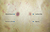
An intradermal test for autoerythrocyte sensitization was performed, with 0.1 mL of the patient's washed red blood cells, whole blood, plasma, and normal saline injected in an inaccessible area of the back, lateral to thoracic spine. Approximately 24 hours later, the patient developed a painful ecchymotic reaction on the washed red blood cell (RBC) and whole blood injection site, while no reaction was observed on the plasma and normal saline injection site (Fig. 2).
Psychiatric evaluation of the patient revealed depression with anxiety features. The patient was empirically given oral antihistamines, steroids and anxiolytics. The ecchymosis reduced and finally diminished approximately 4 weeks later, with a follow-up of stationary phase for more than 3 months.
DISCUSSION
This is a case of AES in an old-aged female. Recurrent episodes of painful, ecchymotic bruising over trunk and extremities were associated with psychiatric problems. The diagnosis was confirmed by inducing similar lesions through the intradermal injection of the patient's own RBC. Correct diagnosis needs careful history taking, knowledge and experiences about AES. Important diagnostic features of AES were described in Table 1.
A large number of pharmacologic agents were tried for the treatment of AES; however, none of these agents proved to be of significant benefit in controlling the manifestations of the disease [5]. Glucocorticosteroids, antibiotics, antihistamines, and antidepressants have been utilized with limited success. In some patients, busulfan, promethasine, amitriptyline, levomepromazine, and hypno- and suggestive therapy as well as psychotherapy may be beneficial. The latter may be especially desirable when there is clear psychogenic provocation [4]. Sometimes a better response to treatment was observed with psychotherapy [6]. In our patient, the lesions did not recur for 3 months after the oral intake of antihistamines, steroids, and anxiolytics empirically.
The prognosis of AES syndrome is good; no fatalities have been reported secondary to the disease. However, the lesions have recurred for as long as 30-40 years. In some cases, the syndrome may remit for months or years and return at a time of severe emotional stress [7].
AES is a rarely reported condition of a still unclear etiology, occurring predominantly in adult females with psychological problems. Stress, hormonal changes and local autoimmune reactions to the patient's own erythrocytes are considered to be the main etiological factors. Groch et al. proposed phosphatidylserine, which could be isolated from the erythrocyte membrane, to be the substance causing the disease. However, patients who were injected with phosphatidylserine showed no reaction [8]. A characteristic feature of the disease is a positive skin reaction to an intradermal injection of the patient's own blood. This condition should be considered in the differential diagnosis of recurrent painful purpuras.
In conclusion, we suggest that the psychosomatic instabilities might have initially disturbed the skin's immunological function and weakened the skin's blood capillaries, so that even a mild trauma of the skin would damage the capillary walls and eventually result in the permeation of RBC. Early diagnosis of AES and early psychiatric support are necessary to avoid later useless interventions.

 XML Download