

 PDF
PDF ePub
ePub Citation
 Print
Print


INTRODUCTION
Pollen grains are important allergen carriers in the environment [1] and are clinically significant to induce IgE-mediated hypersensitivity to at least 40% of allergic individuals [2]. In particular, grass pollen is one of the three main types of allergenic pollens (besides ragweed and trees) [1] that can stimulate pollen-specific IgE reactivity from up to 70-87% among individuals with pollen allergy [3, 4].
The Philippines has a wide range of grass plant species that are releasing large amount of pollen grains throughout the year [5]. Aero-palynological studies had identified the presence of allergenic pollens belonging to the grass family [6-10]. Unfortunately, there are a few serological studies on the allergenicity of these grass pollen grains in the country. For the past 20 years, grass pollen allergen characterization has been reported but most of these studies were conducted in temperate regions where pollen allergy is of major concern. In vitro assays such as sodium dodecyl sulfate polyacrylamide gel electrophoresis (SDS-PAGE) and IgE immunobloting are the primary assessment for this characterization. With the Philippine's allergy prevalence rate of 10% [11] which has also been reported to be continuously increasing [12], it is then necessary to evaluate grass pollen allergy.
The present study reports the sensitization profile of allergic subjects against aqueous extracts of pollen from selected grasses. All the grass species showed IgE reactivity which may explain the allergic symptoms of the subjects. Additionally, IgE-immunoblotting revealed IgE-binding proteins from Saccharum spontaneum, Sporobulus indicus and Choris barbata pollen, which could be further characterized as new allergens.
MATERIALS AND METHODS
Subjects
Using the questionnaire on allergic screening adapted from the International Study of Asthma and Allergies in Childhood [13], individuals with allergic symptoms were recruited in Bayombong, Nueva Vizcaya, Philippines. This area is composed of 17% grassland and 60% agricultural fields [14] and since no allergy profiles were available, the topography and vegetation gave a greater chance to enrol grass pollen allergic subjects.
Three hundred forty one individuals with ages ranging from 2 to 79 years participated in the study. Initial screening was based on the criteria set by the questionnaire [13]. The questionnaire was translated in local dialect and tested for its understandability. It was used as a guide to acquire information on the subjects' symptom which was in the form of an interview. Additional inclusion criteria include being a naturally born Filipino with Filipino parents, have been living in the study base [15], and must not have any history of helminthic infection. As a confirmatory test, total serum IgE level of ≥100 IU/mL [16-22] were conceptually defined as individuals suffering from allergic asthma, rhinitis, dermatitis and conjunctivitis in the study. Using the presented criteria, a total of 141 subjects were qualified and included in the study as allergic cases.
Similar protocols and inclusion criteria were acquired for the non-allergic or the control group. The evaluation of the questionnaire should show that they do not have any allergic symptoms. As for the total serum IgE, they should be <100 IU/mL. Age and gender of the allergic subjects were match-paired with the non-allergic subjects.
The sampling was done from January to March 2009 with permission from the local health office, municipal barangay leaders and hospital administration. Explanatory letter and consent form were provided to all participants.
Collection and isolation of sera
Peripheral blood was extracted by venipuncture from willing subjects. Sera were isolated by centrifugation at 2,500 rpm using a Clay Adams Brand Compact II Centrifuge (Pharmingen, USA). These were stored in 1 mL aliquots at -20℃ until use.
Grass pollen grain collection and extraction
Pollen samples were collected from mature anthers of the grasses as described [23]. Purity of the samples was checked through microscopy (Olympus BX50; Olympus Corp., Japan).
The collected pollen grains were defatted with diethyl-ether (1/10 w/v) and extracted with PBS (0.1 mol/L sodium phosphate containing 0.15 mol/L NaCl, pH 7.3) (1/10 w/v). The pollen with PBS was stirred overnight at 4℃ and centrifuged at 14,000 rpm for 20 min. The supernatant was dialyzed and passed through a 0.22 µm Millipore filter (Millipore Corp., USA). Extracts were stored at -20℃ until use. The total protein content of the extract was analyzed using a Bradford assay [24], with bovine serum albumin as the standard.
SDS-PAGE
Reducing SDS-PAGE of the grass pollen extracts was carried out on 20% polyacrylamide gel using the Mini-PROTEAN II SDS-PAGE Apparatus (BioRad Laboratories, USA). Protein bands in the SDS-PAGE gel were visualized by silver staining (BioRad Laboratories, USA). A combination of broad range and low range proteins markers (Fermentas Life Sciences, Lithuania) was used to assess the proteins.
Total and specific-IgE level determination
Enzyme-linked Immunosorbent Assay (ELISA) was performed to quantify the total and specific-IgE levels of each serum using anti-human IgE biotin-conjugate antibody (Pharmingen, USA) and 4-Nitrophenyl phosphate disodium salt hexahydrate (Sigma-Aldrich, USA) substrate based on the protocols described [25]. The absorbance was measured using the Bio-Tek® ELISA reader (BioTek, USA). Positive specific-IgE levels of the allergic subjects depended on the specific-IgE levels of the non-allergic group on each grass species.
IgE immunoblotting
The extracts were resolved by SDS-PAGE as described above prior to protein transfer onto nitrocellulose membrane. The strips were cut having 0.5 cm width then blocked with 5% non-fat milk. Sera were incubated on the strip overnight at 4℃. Biotinylated anti-human IgE followed by ExtrAvidin (Sigma-Aldrich, USA) and applied with AP conjugate substrate (BioRad Laboratories, USA) was used for the detection.
Statistical analysis
As total and specific IgE levels were high, a logarithmic transformation (Log 10) was used. Statistical computations were performed using Statistical Package for the Social Sciences with a p-value = 0.05. Correlations were made using Spearman's correlation analysis and the difference of the IgE levels between two groups was computed using non-parametric Mann Whitney U test.
RESULTS
Characteristics of subjects
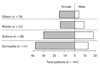
The 141 subjects had an age range of 3 to 79 years, the population had a mean of 26.4 years and 20.9 years standard deviation. Fig. 1 show that 36% of the subjects suffered from dermatitis, 34% had asthma, and 16% had rhinitis. The rest of the subjects were with conjunctivitis and with a combination of 2 or more of the allergic symptoms. Female subjects had higher preponderance than male having a ratio of 2:1 (female:male). No significant values with respect to gender and symptom were present.
Characterization of grass pollen extracts
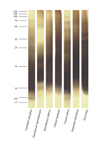
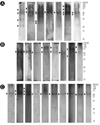
The protein concentration of the extracts ranged from 11 to 22 mg/mL. About 22 mg of total protein was extracted for every gram of pollen grains as a starting material. In the SDS-PAGE (Fig. 2), the extracts resolved bands between 250 to 1.7 kDa. Though some of the proteins appear to be degraded, some bands can be seen at specific levels. Bands from Cynodon dactylon ranged from 72 to 4.6 kDa with distinct bands at 70, 28, 17 to 8, and at 4.6 kDa. Prominent bands can be seen at 60, 20 and 12 kDa for S. spontaneum; 26, 17-20 and 12 kDa for S. indicus; and at 95, and 36 kDa for C. barbata. For Oryza Sativa, it has 95 to 1.7 kDa bands which predominated at the 95, 70, 45, 36 to 28, and 20 to 1.7 kDa levels. Imperata cylindrica and Zea mays bands were equally distributed between the 250 to the 4.6 kDa range.
Total and specific IgE levels
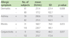
The total IgE levels of the allergic subject had a mean of 209.25 IU/mL (SD = 168.28 IU/mL). With respect to age and sex, no significant relationship was found unlike with the symptoms. Table 1 show significant differences in the total IgE level for the symptoms.
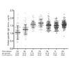
In Fig. 3 the grass specific-IgE levels of the subjects is shown. The subjects' total IgE levels were found to be correlated with their specific-IgE level against S. spontaneum and I. cylindrica with a calculated r of 0.17.
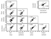
In the analysis of the number of extract, 99% of the subjects have sensitivity to at least one of the grass species. Of which, a high percentage of 83% were reacting to at least 3 pollen extracts suggesting multiple sensitization among the subjects. When correlating the specific-IgE levels of the subjects, Fig. 4 shows that the level within and among species are highly associated. One group involved C. dactylon and S. spontaneum with an r = 0.902 which means they are highly correlated, while the other group involves all other species with correlation values from 0.474 to 0.836.
Specific-IgE level against O. sativa shows a significant difference between subjects having dermatitis. Fifty-seven of the subjects with dermatitis had mean specific-IgE level of 46.05 IU/mL (SD = 33.65) which was found to be significantly different (p = 0.05) with the 73 subjects with no dermatitis and have specific-IgE values of 36.65 IU/mL (SD = 33.59).
IgE immunoblot
Since no reports on the allergenicity of S. spontaneum, S. indicus and C. barbata species were available in allergen databases; further investigation using IgE immunobloting was done. Ten of the most reactive sera from the specific-IgE levels were used to evaluate the extracts.
Fig. 5 shows that S. indicus extract have eight bands that could be recognized by seven out of the ten tested sera. These faint but distinct bands ranged from high to low molecular weights (Fig. 5A). High molecular weight proteins recognized are approximately at 37, 50, 75 and 100 kDa while four lower molecular weight proteins were located between approximately 20 to 25 kDa. For S. spontaneum, 8 out of 10 of the sera showed three IgE reactive protein bands. One band is located at the 90 to 100 kDa molecular weight, another at the 55 kDa and one at 37 kDa (Fig. 5B). Three distinct bands were prominent for C. barbata (Fig. 5C). One, which is at 50 kDa was recognized by all of the 10 subjects. A 70 kDa band was recognized by two of the subjects while one band was shown at approximately 100 kDa.
DISCUSSION
Because of the limited number of studies regarding grass pollen allergy in the Philippines, preliminary assessment of sera from allergic subjects and characterization of natural grass pollen extracts to which these sera show reactivity were presented in this study. The first part of the discussion dealt with the serological characterization of the allergic subjects and next part described the IgE-reactivities detected in the pollen extracts.
As IgE plays a very important role in the mechanisms of allergy [22, 26], both the total and the grass-specific IgE levels were investigated. One of the significant findings is that the serum total IgE level is not associated with either the age or sex of the subjects. This has been supported by the report given by Satwani et al. [22] but contradicting to some studies where higher total IgE levels were more in favour of males in both children [19] and adults [27-29]. Concerning total serum IgE level and the allergic symptoms, subjects with dermatitis have relatively higher total IgE level. This too, was detected from patients suffering from dermatitis [30, 31]. However, this observation should have also been the true for patients having rhinitis and conjunctivitis [19], but strangely, individuals with rhinitis and conjunctivitis in this study have low total IgE levels, similar with the observation done by Mediaty and Neuber [32].
Generally, allergic asthma and allergic rhinitis are the common symptoms for grass pollen allergy [33, 34], although, few report that grass pollen could also elicit allergic dermatitis in atopic patients [35-37]. The high percentage of allergic dermatitis among the subjects could be explained by one of the limitations of this study which is the lack of in vivo assay (e.g. skin prick test). Mainly, the total and grass-specific IgE levels, and IgE immunoblotting were used to assess or confirm the allergic symptoms among the subjects and likewise with the significance of the grass extracts on allergy.
For the characterization of the grass pollen extracts, 14 grass species which were locally available in the area of study were initially selected. Among these grasses were Axonopus compressus, Digitaria sanguinalis, Eleucine indica, Polytrias amaura, Zoysia tenuifolia, Axonopus sp, and Eragrostis tenella. Surprisingly, these species did not show any IgE activity from the allergic subjects' sera even though A. compressus has been reported to have an allergenic potential [38]. On the other hand, C. dactylon, C. barbata, I. cylindrica, S. spontaneum, S. indicus, O. sativa and Z. mays have shown IgE reactivity. Where I. cylindrica, O. sativa, C. dactylon, Z. mays have been reported to contain allergens (www.allergome.com) which may explain the reactivity detected from the sera.
In the analysis of the grass specific-IgE levels of the sera for each extract, significant correlations between and among the grass extracts were detected. Multiple-sensitization among the allergic individuals was also present suggesting that there are similarities of proteins or allergens in the extracts. This similarity or cross-reactive proteins, regardless of biological source, share homologous protein structures eliciting cross-recognition of IgE paratopes [39]. In grasses, cross-reactive allergens include: the grass group 1 allergens (beta-expasins) which is most prominent allergens from grasses detected in 19 species [40]; the grass group 5 allergens, though until now it has no known function was found to be identified in members of Pooidieae subfamily [39, 40]; and lastly, the grass group 12 allergens (profilin), which is an ubiquitous plant allergen [41].
Moreover, the phylogeny also plays an important role to the cross-reactivities among grass species. Only 5 among the 12 subfamilies of grass have some members that are of allergenic significance wherein the majority of these allergenic grass pollen grains belong to the Pooideae subfamily, commonly found in the temperate regions. Other genera which are also considered as an important allergenic species belong to Panicoideae, Chloridoideae, Arundinoideae and Bambusadeae [40]. Unexpectedly, the species which were detected to have IgE reactivity coincide with the mentioned 5 important subfamilies. S. spontaneum, I. cylindrica, Z. mays is a member of the Panicoideae subfamily; C. dactylon, S. indicus, C. barbata belonged to Chloridoideae subfamily; and O. sativa to Arundinoideae subfamily.
Aside from the stated above, cross-reactive carbohydrate determinants (CCD) can also explain the very high IgE reactivity among the sera. These CCDs involve plant and insect glycoproteins [42]. Anti-CCD IgE among allergic patients were found to be mainly reactive to the fucose and xylose residues of these glycans [43], although these IgE were reported to give no clinical significance [39, 44, 45]. Hence, further characterization of both the sera and the extracts is highly recommended.
In the characterization of S. spontaneum, S. indicus and C. barbata extracts, it revealed that only some of the sera reacted on few proteins. This reactivity may be explained by the low IgE titer in the sera and/or amount of the allergen on the extracts. The 50 kDa band identified in all 3 species can be designated as grass group 4 allergens since already reported allergens state that this allergen group have molecular weights between 40-60 KDa [46]. Meanwhile, 20-35 kDa proteins, similar with grass group 5 allergens [46], are also present in S. indicus. The importance of these bands have a worldwide impact because these species can be found in various parts of the world [47-49]. In conclusion, IgE sensitization of allergic subjects in the Philippines may be due to the IgE-binding potential of the proteins present in C. dactylon, S. spontaneum, S. indicus, C. barbata, O. sativa, I. cylindrica, and Z. mays.
 XML Download