

 PDF
PDF ePub
ePub Citation
 Print
Print


INTRODUCTION
Asthma is regarded as a heterogeneous, chronic airway inflammatory disease comprising various phenotypes, and is characterized by reversible airway obstruction and airway hyperresponsiveness (AHR) [1]. Asthma phenotypes can be categorized by clinical features as well as by immunopathology on the basis of inflammation patterns, with regard to the existence or absence of particular inflammatory cell types such as eosinophils or neutrophils [1-3]. The identification of asthma phenotypes has potential clinical importance, because natural history, disease prognosis, and treatment response may differ according to phenotype [1, 2].
Eosinophils have long been believed to play a central role in asthmatic airway inflammation [4]. Early biopsy studies revealed eosinophilic inflammation within the airway mucosa of asthmatic patients [5]. The functional relevance of eosinophils is supported by studies demonstrating that eosinophilic airway inflammation was improved after inhaled corticosteroid treatment [6], was aggravated by allergen exposure [7], and was correlated with asthma severity [8]. However, as these studies were performed in small populations, they afford a limited representation of the general asthmatic population.
The development of simple, noninvasive methods for assessing airway inflammation using induced sputum has provided a way of studying airway inflammation in a diverse range of patients [9, 10]. In many studies using these methods, sputum eosinophilia was not observed in a significant proportion of asthma patients [11-13]. Sputum eosinophilia was not detected in as many as 25% of untreated symptomatic patients [11], patients with severe refractory asthma [13], and patients experiencing asthma exacerbation [12], and in up to 50% of patients taking high-dose inhaled steroids [14]. Noneosinophilic asthma (NEA) has been estimated to be present in 49% of patients with symptomatic asthma presenting to a respiratory clinic [15]. Furthermore, the absence of sputum eosinophilia was reported to be a stable feature in a number of patients over a 12-month period in a longitudinal study [16]. Based on these findings, recent reports have suggested the importance of NEA as a distinct phenotype of asthma [15, 17].
Studies have suggested that NEA is associated with increased neutrophil and interleukin (IL)-8 levels and that non-allergic, neutrophil-driven airway inflammation is the underlying mechanism for NEA [15]. The activation of an innate immune response provoked by endotoxins, viral or bacterial infections, cigarette smoke, air pollutants, or occupational agents has been linked with the pathogenesis of noneosinophilic, neutrophilic inflammation [15, 17]. From a clinical point of view, NEA is associated with smoking, obesity, and a poor response to an inhaled corticosteroid (ICS) [15, 17]. Although many studies have shown that low sputum eosinophil counts are associated with poor outcomes of ICS treatment, most were performed with short treatment durations and in limited populations [17]. To date, there is little information regarding the long-term treatment outcome of NEA. In addition, the clinical significance of the absence of sputum eosinophilia has not been fully explored in Korean symptomatic asthmatics. The purpose of the present study was to evaluate clinical significance of sputum eosinophilia and long-term treatment responses of Korean asthmatics with and without sputum eosinophilia.
MATERIALS AND METHODS
Subjects
A total of 201 asthma patients who had undergone induced sputum analysis before regular asthma treatment were enrolled in this study. All were recruited from the previously established Cohort for Reality and Evolution of Adult Asthma (COREA) [18] according to the following inclusion criteria: (1) airway reversibility documented by an increase of forced expiratory volume in one second (FEV1) of 12% and 200 mL after inhalation of 200 µg of salbutamol, or airway hyperresponsiveness documented by a methacholine provocation concentration causing a 20% fall in FEV1 (PC20) of ≤16 mg/mL, in the past or at the time of enrollment; (2) presence of persistent characteristic asthma symptoms and physician's diagnosis of asthma; (3) availability of adequate induced sputum analysis data at the time of enrollment; and (4) absence of asthma controller medication, including systemic ICS, leukotriene antagonists, and theophylline, for at least 3 months before enrollment and induced sputum analysis. Of the 201 subjects, 53 com-pleted 24 months of regular follow-up visits, and the analysis of long-term treatment outcomes was performed with this group. All subjects were provided written informed consent, and the study protocol was approved by the institutional review board of each participating hospital.
Study design
Clinical evaluation, spirometry, allergy skin-prick tests, a methacholine bronchial provocation test, blood tests, chest and paranasal sinus simple radiological exams, and sputum eosinophil analysis were performed initially at the time of enrollment. The subjects received treatment based on symptoms and lung func tion, according to the recommendations of the Global Initiative for Asthma [19]. Asthma symptom scores, presence of exacerbation, and treatment compliance were evaluated every 3 months during the 24-month follow-up period. Lung function was evaluated at 6-month intervals, using spirometry equipment and techniques that conformed to the criteria issued by the American Thoracic Society and European Respiratory Society [20]. Skin-prick tests were performed using 11 inhalant allergens (Bencard, UK; or Aller-gopharma, Germany), including Dermatophagoides pteronyssinus, D. farinae, tree pollen mixture, grass pollen mixture, ragweed, mugwort, cockroach, Alternaria, Aspergillus, cat, and dog. Wheal development 15 min later was used to assess positive responses to allergens. Atopy was defined as a positive skin-prick test response (allergen/histamine ratio ≥1 and mean wheal size >3 mm) to one or more allergens.
Induced sputum analysis
Sputum induction and processing were carried out according to a standardized protocol, as previously described [21]. Briefly, after measurement of basal FEV1, all study participants were pretreated with 200 µg of salbutamol (Ventolin™; GlaxoSmithKline, England). To induce sputum production, a 4.5% hypertonic saline solution was administered for four times at 5-min intervals using an ultrasonic nebulizer (Omron, Japan) with the output set at 4.5 mL/min. The patients rinsed their mouths and carefully spit the sputum into a Petri dish. The more viscid and denser portions were selected and processed. After the addition of dithiothreitol (DTE, 0.01 M), the sputum samples were mixed by rotation for 20 min at room temperature and filtered through 52-mm nylon gauze to remove debris and mucus. The cell pellets were collected by centrifugation at 450 × g for 10 min and resuspended in phosphate-buffered saline to a volume equal to that of the original sputum sample plus DTE. The cells were counted using a hemacytometer, and cell concentrations were adjusted to 1.0 × 10⁶ cells/mL. Cytospins were prepared by adding 60 µL of this cell suspension to Shandon II cytocentrifuge cups (Shandon Southern Instruments, USA) and were spun for 5 min at 42 × g. Slides were prepared, and the cells were stained with Diff Quik solution (Sysmex, Japan). To determine cell differentiation, 300 nucleated cells per slide were counted, and macrophage, lymphocyte, neutrophil, and eosinophil values were calculated as percentages of total inflammatory cells, excluding squamous epithelial cells. Sputum samples that contained more than 30% squamous epithelial cells were not analyzed. Subjects were classified as either NEA or eosinophilic asthma (EA) based on the initial sputum eosinophil count. NEA was defined as asthma with sputum eosinophil counts <3%, and EA was defined as asthma with sputum eosinophil counts of ≥3% [22].
Statistical analysis
All statistical analyses were performed using SPSS ver. 16.0 (SPSS, USA). Categorical data were compared using a chi-squared or Fisher's exact test. Numerical data were compared using Student's t-test or a Mann-Whitney test. Linear regression was applied to account for the effects of age, gender, body mass index (BMI), and smoking on changes in the pulmonary function test results. Differences in FEV1 and symptom scores over time within groups were assessed with a Wilcoxon matched-pair t-test. A value of p < 0.05 indicated statistical significance.
RESULTS
Clinical characteristics of 201 steroid-naive asthmatics
Of the 201 asthmatic patients included in the study, 97 (48.3%) were in the NEA group and 104 (51.7%) were in the EA group. Mean age was 51.2 years and 44.4% was male.
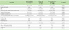
Of the 201 subjects, 53 completed 24 months of regular follow-up visits and 148 did not complete the study. But no difference in age, gender, BMI or smoking habit was found between the followed-up patients and followed-up loss patients. There was no difference in the baseline FEV1, symptom duration, asthma severity, or prevalence of atopy, or allergic rhinosinusitis between the two groups (Table 1).
Relationship between sputum eosinophil/neutrophil profiles and lung function and airway hyperresponsiveness in steroid-naive asthmatics
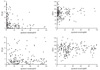
We evaluated the correlation between sputum inflammatory cell profiles and lung function and airway hyperresponsiveness in the steroid-naive state. Correlation analyses were performed using percentages of eosinophils and neutrophils in the induced sputum collected at baseline (Fig. 1). A higher percentage of sputum eosinophils was associated with a lower PC20 (r = -0.193; p = 0.009, Spearman correlation), but not with FEV1 (r = 0.045; p = 0.525). The percentage of sputum neutrophils was not associated with either PC20 (r = 0.104, p = 0.143) or FEV1 (r = 0.003, p = 0.973).
Clinical characteristics of NEA and EA in steroid-naive asthmatics who completed 24 months of follow-up visits
A total of 53 patients (22 NEA and 31 EA patients) who completed 24 months of follow-up visits were analyzed to compare long-term asthma treatment responses between the NEA and EA groups. No difference in age, gender, BMI or smoking habit was found between the NEA and EA groups. Baseline blood eosinophil counts were significantly lower and baseline blood neutrophil counts were significantly higher in the NEA group than in the EA group. There was no difference in the baseline FEV1 and FVC between the two groups and there was no significant difference in symptom duration, asthma severity, or allergic rhinosinusitis between the NEA and EA groups. Initial asthma medication use, including ICSs, long-acting β2-agonists, leukotriene antagonists, theophylline, and oral corticosteroids, was also similar between the two groups (Table 2).
Sputum differential cell counts within the total non-squamous cell count
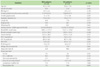
As expected, the mean percentage of eosinophils (as the percentage of the total number of nonsquamous cells) in NEA patients was significantly lower than EA patients (0.6 ± 0.8 vs. 27.5 ± 26.5, p < 0.0001). No difference in sputum level of the mean percentage of neutrophils was found between NEA patients and EA patients (Table 3).
FEV1 improvement in NEA and EA patients during the 24-month follow-up period
Throughout the 24-month follow-up period, the improvement in FEV1 was less in NEA patients compared with EA patients, although the difference was statistically significant only at 12 (0.7 ± 11.1 vs. 25.3 ± 56.7, p = 0.048) and 18 months (3.1 ± 11.6 vs. 18.3 ± 29.6, p = 0.021). However, after adjusting for the effects of demographic factors such as age, gender, BMI, and smoking habit, the improvement in FEV1 was significantly less in the NEA group than in the EA group at every 6-month visit during the 24-month follow-up (Table 4).
Changes in FEV1 over time in NEA and EA patients
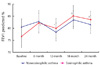
We analyzed FEV1 of total of 53 patients (22 NEA and 31 EA patients) who completed 24 months of follow-up visits. The changes in FEV1 and symptom scores over time in each group were assessed using a Wilcoxon matched-pair t-test. EA patients showed significantly improved FEV1 at 6 months of treatment compared with the baseline FEV1 (82.2 ± 20.3% vs. 76.6 ± 25.2%, p = 0.002), and this improvement was sustained at 12, 18, and 24 months (80.0 ± 20.2, 85.1 ± 22.6, and 83.6 ± 19.7%, respectively; p < 0.05). However, there was no improvement in FEV1 in NEA patients at any 6-month follow-up visit (82.9 ± 18.3, 78.5 ± 16.5, 83.5 ± 16.2, 82.2 ± 18.3, and 81.6 ± 17.1%, respectively; p > 0.05) (Fig. 2). Symptom scores improved in both groups, with no significant difference between the groups. The frequency of acute exacerbation was not significantly different between the groups (data not shown).
DISCUSSION
Our observational study showed that NEA patients had no significant lung function improvement during 24 months of guideline-based regular controller treatment. In contrast, EA patients experienced significant long-term FEV1 improvement and better treatment outcomes compared with NEA patients.
Airway inflammation is the basis of asthma pathogenesis, and the goal of asthma treatment using ICSs is the targeted control of airway inflammation for optimal lung function and symptom control [19]. However, given the heterogeneous inflammatory processes involved in the pathogenesis of asthma, the clinical features and treatment responses of asthmatics can vary according to individual underlying inflammatory or immunological phenotypes [1, 2]. EA is well established as classical allergic asthma mediated by Th2 cytokines, including IL-4, IL-5, and IL-13, that orchestrate eosinophilic airway inflammation and IgE production [3]. An increased sputum eosinophil percentage has been reported to be associated with more severe asthma, more severe airflow limitation, and increased airway hyperresponsiveness [8, 23, 24]. Nevertheless, studies using induced sputum analysis have revealed that not all patients with severe asthma have airway eosinophilia and that the noneosinophilic asthma phenotype is present across all grades of severity [13, 25].
In the present study, we observed that only 52% of steroid-naive asthmatic subjects had elevated baseline sputum eosinophil levels. A higher sputum eosinophil percentage was correlated with a higher methacholine-induced airway hyperresponsiveness (lower PC20), but not with lung function (prebronchodilator FEV1). Sputum neutrophil percentage was not correlated with methacholine PC20 or prebronchodilator FEV1. In accordance with previous study results showing that AHR is associated with sputum eosinophil, but not sputum neutrophil levels [24], our findings suggest that sputum eosinophils are important mediators of AHR. However, an association between the numbers of sputum inflammatory cells (both eosinophils and neutrophils) and airway obstruction, which has been reported by several investigators [24, 26], was not found in our study population.
Most previous studies have shown that NEA is relatively steroid-unresponsive and that sputum eosinophils are markers for a response to ICSs or systemic steroid therapy in asthmatics [17]. Several clinical trials have compared the treatment response to corticosteroids between steroid-naive NEA and EA patients. Green et al. [11] reported that compared with EA patients, NEA patients had significantly less improvement in FEV1, PC20, and visual analogue symptom scores after 2 months of inhaled budesonide treatment. A study by Bacci et al. [27] showed that low sputum eosinophil counts were associated with a lack of FEV1 improvement following 2-4 weeks of treatment with inhaled beclomethasone.
In a 2-week oral steroid trial in chronic stable asthmatics, Little et al. [28] found that sputum eosinophilia had a positive predictive value of 64% and negative predictive value of 68% (sensitivity 54%, specificity 76%) for an increase in FEV1 of ≥15% after treatment. Moreover, Meijer et al. [29] observed that sputum eosinophilia predicted the FEV1 response as well as PC20 and quality of life improvement after 2 weeks of corticosteroid treatment.
Unlike previous studies using short courses of inhaled or systemic steroid treatment, our study was a 24-month, long-term observational study and showed no significant improvement in FEV1 in NEA patients. This study was not conducted with a fixed medication regimen over the study period; instead, our subjects were given controller medications, including at least an ICS, and were followed with adjustments of medications according to the guidelines. The medication pattern was similar between the EA and NEA groups. Despite guideline-based therapy, the NEA patients showed no significant lung function change at any of the follow-up visits, whereas EA patients exhibited significantly improved FEV1 within 6 months, and the improvement was sustained for 24 months. These results suggest that sputum eosinophilia may be a useful predictor of long-term response to asthma therapy and that current asthma controller medications, including ICSs, long-acting β2-agonists, and leukotriene antagonists, are less effective in patients with NEA than in those with EA.
The pathogenesis of NEA involves the activation of an innate immune response and neutrophilic airway inflammation that generally tends to be unresponsive to steroids [15, 17, 30]. Environmental exposure to bacterial endotoxins, viruses, particulate air pollution, or ozone is an important trigger for neutrophilic airway inflammation [15]. Proinflammatory cytokines, including IL-1, IL-8, and tumor necrosis factor a, together with Toll-like receptor and CD14 contribute to the development of noneosinophilic, neutrophilic inflammation in response to the activation of innate immunity [15, 17]. Recent reports have indicated that Th17 cells evoke neutrophilic airway inflammation and that Th17 cell-mediated airway inflammation displays a more severe, steroid-resistant asthma phenotype [31, 33]. Our finding that NEA failed to show an improvement in FEV1 despite appropriate 24-month controller treatment is likely to be associated with the poor steroid response of neutrophilic airway inflammation mediated by IL-8, tumor necrosis factor a, or Th17 cells.
There are several limitations to our study. First, the size of the study population that completed the 24-month follow-up visits was small, possibly because the study subjects originated from an existing observational adult asthma cohort. Selection bias might have affected our results, although there were no significant differences in demographic and clinical characteristics between the follow-up and non-follow-up groups. Larger prospective studies are needed to corroborate these findings. Second, the study was not conducted with a fixed medication regimen, as discussed above. Third, the analysis of sputum eosinophilia was performed only once, at the baseline point of study entry, and was not repeated. Thus, the diagnosis of NEA or EA was based on a single sample of induced sputum. However, a recent study has indicated that a cut-off point of 3% eosinophils allows the presence of eosinophilia to be reliably determined from a single sputum sample [22].
Despite these limitations, our findings provide useful insights into the heterogeneity of asthma and its response to therapy. NEA patients showed poor lung function improvement compared with EA patients during the 24 months of follow-up in our Korean asthma cohort population. Thus, sputum eosinophils may predict the response to therapy, and NEA as a distinct clinical phenotype may relate to poor long-term treatment outcomes.


 XML Download