

 PDF
PDF ePub
ePub Citation
 Print
Print


Introduction
Naringenin, a colorless flavanone present in a variety of herbs and fruits including grapefruit and tomatoes, has been found to be highly beneficial to human health due to its strong antioxidant potential.1 It was reported that naringenin has anti-inflammatory, antiproliferative, anticancer, chemopreventive and estrogenic properties,234 and has beneficial influence on lipid metabolism and insulin sensitivity.5 The pharmacological application of naringenin-type flavonoids has become more attractive due to the possibility of the chemical synthesis of its prenylated derivatives.6
(±)-6-(1,1-Dimethylallyl)naringenin (6-DMAN, 1), a natural naringenin-type flavonoid isolated originally from the leaves of the African tree Monotes engleri (family Dipterocarpaceae),7 has been found to exhibit estrogenic and antiandrogenic properties,38 and also found to display cytotoxic activity against several human cancer cell lines.7 (±)-5-(O-Prenyl)naringenin-4′,7-diacetate (5-O-PN, 2), a synthetic derivative of 6-DMAN, showed high cytotoxicity in HL-60 and MCF-7 cell lines.9
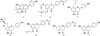
No metabolism study has yet been carried out to identify the metabolic fate of 6-DMAN and 5-O-PN. Hence, metabolic processes of 1 and 2 were investigated by using microorganisms in the present study. Biotransformation studies using microorganisms are well known as an important tool for the conversion of natural compounds. It has been used successfully as in vitro models to mimic and predict the metabolic fate of pharmaceutical agents in mammalian systems.101112 Scale-up studies with three microbial cultures, Mucor hiemalis, Cunninghamella elegans var. elegans, and Penicillium chrysogenum, have resulted in the production of five microbial metabolites (Fig. 1).
Experimental
General experimental procedures
The 1H and 13C NMR spectra were obtained in CDCl3 and DMSO-d6 on a Varian Unity Inova 300 spectrometer at 300 and 75 MHz, respectively. The chemical shift values (δ) are reported in ppm units, and the coupling constants (J) are in Hz. UV spectra were recorded on a JASCO V-530 spectrophotometer, and IR spectra were obtained on a JASCO FT/IR-300E spectrometer in KBr discs. Optical rotations were measured with a Perkin Elmer 343 Plus polarimeter. ESIMS and HRESIMS analyses were performed on a Micromass QTQF2 mass spectrometer. TLC analyses were carried out on precoated silica gel 60 F254 plates (Merck, Darmstadt, Germany). The developing system used was chloroform:methanol (9:1, v/v) solution, and visualization of the TLC plates was performed using anisaldehyde-H2SO4 spray reagent. For column chromatography, the adsorbent used was silica gel 60 (70 - 230 mesh, Merck). HPLC was performed on a Waters 600E Multisolvent Delivery System (Waters Corp., Milford, MA, USA) connected to a Waters 486 detector using Phenomenex C18 column (10 × 250 mm, 5 µm) with MeOH:H2O at a flow rate of 2.0 mL/min.
Materials and microorganisms
6-DMAN (1) and 5-O-PN (2) were semi-synthesized from rac-naringenin.13 All of the ingredients for microbial media, including dextrose, peptone, malt extract, yeast extract, and potato dextrose broth were purchased from Becton, Dickinson and Company (Sparks, MD, USA).
All the microorganisms were obtained from the Korean Collection for Type Cultures (KCTC). Twenty-four cultures were used for the preliminary screening process and are listed below: Absidia coerulea 6936, Alternaria alternata 6005, Aspergillus fumigatus 6145, Candida famata 7000, Cunninghamella elegans var. elegans 6992, Debaryomyces hansenii var. hansenii 7645, Debaryomyces occidentalis var. occidentalis 7194, Filobasidium neoformans 7902, Fusarium merismoides 6153, Gliocladium deliquescens 6173, Glomerella cingulata 6075, Hormoconis resinae 6966, Kluyveromyces marxianus 7155, Metschnikowia pulcherrima 7605, Monascus rubber 6122, Mortierella ramanniana var. angulispora 6137, Mucor hiemalis 26779, Penicillium chrysogenum 6933, Pichia pastoris 7190, Rhizopus oryzae 6946, Saccharomycodes ludwigii 7126, Torulaspora delbrueckii 7116, Trichoderma koningii 6042, Trigonopsis variabilis 7263.
Three types of media were used in the fermentation experiments and are listed below: A. coerulea and M. hiemalis were cultured on malt medium (malt extract 20 g/L, dextrose 20 g/L, peptone 1 g/L); C. elegans var. elegans was cultured on potato dextrose medium (24 g/L); other microorganisms were cultured on yeast-malt medium (dextrose 10 g/L, peptone 5 g/L, malt extract 3 g/L, and yeast extract 3 g/L).
Screening procedures
Microbial metabolism studies were carried out according to the standard two-stage procedure.10 Briefly, the actively growing microbial cultures were inoculated in 100 mL Erlenmeyer flasks containing 25 mL of a suitable medium, and incubated with gentle agitation (200 rpm) at 25 ℃ in a temperature-controlled shaking incubator. The DMSO solutions (10 mg/mL, 100 µL) of 1 and 2 were added to each flask 24 h after inoculation, and further incubated at the same conditions for another 8 days. Sampling and TLC monitoring were performed at an interval of 24 h. Culture controls consisted of fermentation cultures in which the microorganisms were grown without addition of substrates.
Scale-up fermentations of 6-DMAN and 5-O-PN
Scale-up fermentations were carried out under the same temperature-controlled shaking conditions with three or eight 500 mL Erlenmeyer flasks each containing 125 mL of a suitable medium, and 45 mg of 1 and 100 mg of 2 dissolved in DMSO were distributed evenly among flasks, respectively. After incubation for 7 days, the microbial culture broth was extracted with EtOAc (400 mL × 3), and the organic layers were combined and concentrated in vacuo. The EtOAc extract (80 mg) of 1 from M. hiemalis culture broth was chromatographed by semi-preparative reversed-phase HPLC with 80% MeOH as mobile phase to give metabolite 3 (3.0 mg). The EtOAc extract (70 mg) of 1 from C. elegans var. elegans culture broth was chromatographed by semi-preparative reversed-phase HPLC with 80% MeOH to give metabolite 4 (4.4 mg). The EtOAc extract (200mg) of 2 from P. chrysogenum culture broth was chromatographed by semi-preparative reversed-phase HPLC with 65% MeOH to afford metabolites 5 (4.5 mg), 6 (1.5 mg), and 7 (3.0 mg).
(±)-6-(1,1-Dimethylallyl)naringenin (1)
A white solid; 1H-NMR (CDCl3, 300 MHz) δ 13.11 (1H, s, 5-OH), 7.48 (1H, s, 7-OH), 7.31 (2H, d, J = 8.9 Hz, H-2′/6′), 6.86 (2H, d, J = 8.9 Hz, H-3′/5′), 6.44 (1H, dd, J = 10.6, 17.9 Hz, H-2″), 5.93 (1H, s, H-8), 5.43 (1H, dd, J = 0.8, 17.9 Hz, H-3″a), 5.36 (1H, dd, J = 0.8, 10.6 Hz, H-3″b), 5.31 (1H, dd, J = 3.1, 12.7 Hz, H-2), 5.10 (1H, s, 4′-OH), 3.07 (1H, dd, J = 12.2, 17.0 Hz, H-3a), 2.78 (1H, dd, J = 3.1, 17.0 Hz, H-3b), 1.60 (3H, s, H-4″), 1.57 (3H, s, H-5″); 13C NMR (CDCl3, 75 MHz) δ 196.2 (C-4), 164.5 (C-7), 163.9 (C-5), 160.4 (C-9), 156.0 (C-4′), 149.6 (C-2″), 130.7 (C-1′), 127.4 (C-2′/6′), 115.6 (C-3′/5′), 113.5 (C-3″), 111.5 (C-6), 103.0 (C-10), 96.7 (C-8), 78.4 (C-2), 43.3 (C-3), 40.7 (C-1″), 27.2 (C-4″), 26.6 (C-5″).
(±)-5-(O-Prenyl)naringenin-4′,7-diacetate (2)
A white solid; 1H-NMR (CDCl3, 300 MHz) δ 7.46 (2H, d, J = 8.4 Hz, H-2′/6′), 7.14 (2H, d, J = 8.4 Hz, H-3′/5′), 6.42 (1H, d, J = 2.0 Hz, H-6), 6.32 (1H, d, J = 2.0 Hz, H-8), 5.53 (1H, t, J = 6.5 Hz, H-2″), 5.43 (1H, dd, J = 2.7, 12.5 Hz, H-2), 4.62 (2H, d, J = 6.5 Hz, H-1″), 3.01 (1H, dd, J = 12.5, 16.9 Hz, H-3a), 2.82 (1H, dd, J = 2.7, 16.9 Hz, H-3b), 2.31 (3H, s, 7-OAc), 2.30 (3H, s, 4′-OAc), 1.79 (3H, s, H-4″), 1.74 (3H, s, H-5″).
(±)-8-Prenylnaringenin (3)
A white solid;  : 0° (c 0.028, MeOH); 1H-NMR (DMSO-d6, 300 MHz) δ 12.18 (1H, s, 5-OH), 10.78 (1H, s, 7-OH), 9.69 (1H, s, 4′-OH), 7.30 (2H, d, J = 8.5 Hz, H-2′/6′), 6.76 (2H, d, J = 8.5 Hz, H-3′/5′), 5.96 (1H, s, H-6), 5.42 (1H, dd, J = 2.8, 12.5 Hz, H-2), 5.08 (1H, t, J = 6.2 Hz, H-2″), 3.20 (1H, dd, J = 12.5, 16.3 Hz, H-3a), 3.08 (2H, d, J = 6.2 Hz, H-1″), 2.71 (1H, dd, J = 2.8, 16.3 Hz, H-3b), 1.58 (3H, s, H-4″), 1.53 (3H, s, H-5″); 13C NMR (DMSO-d6, 75 MHz) δ 196.7 (C-4), 164.3 (C-9), 161.1 (C-5), 159.7 (C-7), 157.5 (C-4′), 130.1 (C-3″), 129.2 (C-1′), 128.0 (C-2′/6′), 122.6 (C-2″), 115.1 (C-3′/5′), 106.9 (C-8), 101.7 (C-10), 95.2 (C-6), 78.2 (C-2), 41.9 (C-3), 25.5 (C-5″), 21.2 (C-1″), 17.6 (C-4″).
: 0° (c 0.028, MeOH); 1H-NMR (DMSO-d6, 300 MHz) δ 12.18 (1H, s, 5-OH), 10.78 (1H, s, 7-OH), 9.69 (1H, s, 4′-OH), 7.30 (2H, d, J = 8.5 Hz, H-2′/6′), 6.76 (2H, d, J = 8.5 Hz, H-3′/5′), 5.96 (1H, s, H-6), 5.42 (1H, dd, J = 2.8, 12.5 Hz, H-2), 5.08 (1H, t, J = 6.2 Hz, H-2″), 3.20 (1H, dd, J = 12.5, 16.3 Hz, H-3a), 3.08 (2H, d, J = 6.2 Hz, H-1″), 2.71 (1H, dd, J = 2.8, 16.3 Hz, H-3b), 1.58 (3H, s, H-4″), 1.53 (3H, s, H-5″); 13C NMR (DMSO-d6, 75 MHz) δ 196.7 (C-4), 164.3 (C-9), 161.1 (C-5), 159.7 (C-7), 157.5 (C-4′), 130.1 (C-3″), 129.2 (C-1′), 128.0 (C-2′/6′), 122.6 (C-2″), 115.1 (C-3′/5′), 106.9 (C-8), 101.7 (C-10), 95.2 (C-6), 78.2 (C-2), 41.9 (C-3), 25.5 (C-5″), 21.2 (C-1″), 17.6 (C-4″).(2S)-5,4′-Dihydroxy-7,8-[(R)-2-(1-hydroxy-1-methylethyl) -2,3-dihydrofurano]flavanone (4)
A pale yellow amorphous powder; : −120° (c 0.056, MeOH); 1H-NMR (DMSO-d6, 300 MHz) δ 12.43 (1H, s, 5-OH), 9.69 (1H, s, 4′-OH), 7.32 (2H, d, J = 8.5 Hz, H-2′/6′), 6.80 (2H, d, J = 8.5 Hz, H-3′/5′), 5.96 (1H, s, H-6), 5.49 (1H, dd, J = 3.0, 12.2 Hz, H-2), 4.74 (1H, t, J = 8.5 Hz, H-2″), 3.26 (1H, dd, J = 12.2, 17.3 Hz, H-3a), 2.90 (2H, dd, J = 3.5, 8.5 Hz, H-1″), 2.72 (1H, dd, J = 3.0, 17.3 Hz, H-3b), 1.11 (3H, s, H-4″), 1.08 (3H, s, H-5″); 13C NMR (DMSO-d6, 75 MHz) δ 196.6 (C-4), 168.9 (C-7), 164.5 (C-5), 158.2 (C-4′), 157.4 (C-9), 129.2 (C-1′), 128.7 (C-2′/6′), 115.6 (C-3′/5′), 105.2 (C-8), 102.7 (C-10), 92.0 (C-2″), 90.9 (C-6), 78.9 (C-2), 70.4 (C-3″), 42.4 (C-3), 26.5 (C-1″), 26.1 (C-5″), 25.5 (C-4″).
: −120° (c 0.056, MeOH); 1H-NMR (DMSO-d6, 300 MHz) δ 12.43 (1H, s, 5-OH), 9.69 (1H, s, 4′-OH), 7.32 (2H, d, J = 8.5 Hz, H-2′/6′), 6.80 (2H, d, J = 8.5 Hz, H-3′/5′), 5.96 (1H, s, H-6), 5.49 (1H, dd, J = 3.0, 12.2 Hz, H-2), 4.74 (1H, t, J = 8.5 Hz, H-2″), 3.26 (1H, dd, J = 12.2, 17.3 Hz, H-3a), 2.90 (2H, dd, J = 3.5, 8.5 Hz, H-1″), 2.72 (1H, dd, J = 3.0, 17.3 Hz, H-3b), 1.11 (3H, s, H-4″), 1.08 (3H, s, H-5″); 13C NMR (DMSO-d6, 75 MHz) δ 196.6 (C-4), 168.9 (C-7), 164.5 (C-5), 158.2 (C-4′), 157.4 (C-9), 129.2 (C-1′), 128.7 (C-2′/6′), 115.6 (C-3′/5′), 105.2 (C-8), 102.7 (C-10), 92.0 (C-2″), 90.9 (C-6), 78.9 (C-2), 70.4 (C-3″), 42.4 (C-3), 26.5 (C-1″), 26.1 (C-5″), 25.5 (C-4″).(±)-5-(O-Prenylnaringenin)-4′-acetate (5)
A pale yellow amorphous powder; : 0° (c 0.052, MeOH); UV (MeOH) λmax: 286, 343 nm; IR (KBr) νmax cm−1: 3345, 1688, 1620, 1596, 1443, 1372, 1208; 1H-NMR (DMSO-d6, 300 MHz) δ 7.53 (2H, d, J = 8.5 Hz, H-2′/6′), 7.16 (2H, d, J = 8.5 Hz, H-3′/5′), 6.04 (1H, d, J = 1.6 Hz, H-6), 5.95 (1H, d, J = 1.6 Hz, H-8), 5.48 (1H, dd, J = 2.8, 12.5 Hz, H-2), 5.39 (1H, m, H-2″), 4.50 (2H, d, J = 6.2 Hz, H-1″), 2.97 (1H, dd, J = 12.5, 16.3 Hz, H-3a), 2.60 (1H, dd, J = 2.8, 16.3 Hz, H-3b), 2.27 (3H, s, -OAc), 1.74 (3H, s, H-4″), 1.69 (3H, s, H-5″); 13C NMR (DMSO-d6, 75 MHz) δ 187.0 (C-4), 169.2 (C-1‴), 165.6 (C-7), 164.0 (C-9), 161.3 (C-5), 150.3 (C-4′), 136.8 (C-3″), 136.4 (C-1′), 127.7 (C-2′/6′), 121.9 (C-3′/5′), 119.9 (C-2″), 104.2 (C-10), 95.8 (C-6), 94.9 (C-8), 77.4 (C-2), 65.1 (C-1″), 44.9 (C-3), 25.5 (C-4″), 20.9 (C-2‴), 18.1 (C-5″); HR-ESI-MS m/z 383.1494 [M+H]+ (calcd for C22H23O6, 383.1495).
: 0° (c 0.052, MeOH); UV (MeOH) λmax: 286, 343 nm; IR (KBr) νmax cm−1: 3345, 1688, 1620, 1596, 1443, 1372, 1208; 1H-NMR (DMSO-d6, 300 MHz) δ 7.53 (2H, d, J = 8.5 Hz, H-2′/6′), 7.16 (2H, d, J = 8.5 Hz, H-3′/5′), 6.04 (1H, d, J = 1.6 Hz, H-6), 5.95 (1H, d, J = 1.6 Hz, H-8), 5.48 (1H, dd, J = 2.8, 12.5 Hz, H-2), 5.39 (1H, m, H-2″), 4.50 (2H, d, J = 6.2 Hz, H-1″), 2.97 (1H, dd, J = 12.5, 16.3 Hz, H-3a), 2.60 (1H, dd, J = 2.8, 16.3 Hz, H-3b), 2.27 (3H, s, -OAc), 1.74 (3H, s, H-4″), 1.69 (3H, s, H-5″); 13C NMR (DMSO-d6, 75 MHz) δ 187.0 (C-4), 169.2 (C-1‴), 165.6 (C-7), 164.0 (C-9), 161.3 (C-5), 150.3 (C-4′), 136.8 (C-3″), 136.4 (C-1′), 127.7 (C-2′/6′), 121.9 (C-3′/5′), 119.9 (C-2″), 104.2 (C-10), 95.8 (C-6), 94.9 (C-8), 77.4 (C-2), 65.1 (C-1″), 44.9 (C-3), 25.5 (C-4″), 20.9 (C-2‴), 18.1 (C-5″); HR-ESI-MS m/z 383.1494 [M+H]+ (calcd for C22H23O6, 383.1495).(±)-Naringenin-4′-acetate (6)
A pale yellow amorphous powder; : 0° (c 0.024, MeOH); 1H-NMR (DMSO-d6, 300 MHz) δ 12.16 (1H, s, 5-OH), 7.55 (2H, d, J = 8.6 Hz, H-2′/6′), 7.19 (2H, d, J = 8.6 Hz, H-3′/5′), 5.87 (1H, d, J = 1.2 Hz, H-6), 5.84 (2H, d, J = 1.2 Hz, H-8), 5.56 (1H, dd, J = 2.7, 13.0 Hz, H-2), 3.26 (1H, dd, J = 13.0, 17.2 Hz, H-3a), 2.74 (1H, dd, J = 2.7, 17.2 Hz, H-3b), 2.29 (3H, s, -OAc).
: 0° (c 0.024, MeOH); 1H-NMR (DMSO-d6, 300 MHz) δ 12.16 (1H, s, 5-OH), 7.55 (2H, d, J = 8.6 Hz, H-2′/6′), 7.19 (2H, d, J = 8.6 Hz, H-3′/5′), 5.87 (1H, d, J = 1.2 Hz, H-6), 5.84 (2H, d, J = 1.2 Hz, H-8), 5.56 (1H, dd, J = 2.7, 13.0 Hz, H-2), 3.26 (1H, dd, J = 13.0, 17.2 Hz, H-3a), 2.74 (1H, dd, J = 2.7, 17.2 Hz, H-3b), 2.29 (3H, s, -OAc).(±)-Naringenin (7)
A white solid; : 0° (c 0.021, MeOH); 1H-NMR (DMSO-d6, 300MHz) δ 12.17 (1H, s, 5-OH), 7.30 (2H, d, J = 8.5 Hz, H-2′/6′), 6.80 (2H, d, J = 8.5 Hz, H-3′/5′), 5.86 (2H, s, H-6/8), 5.41 (1H, dd, J = 2.8, 13.0 Hz, H-2), 3.25 (1H, dd, J = 13.0, 17.0 Hz, H-3a), 2.68 (1H, dd, J = 2.8, 17.0 Hz, H-3b).
: 0° (c 0.021, MeOH); 1H-NMR (DMSO-d6, 300MHz) δ 12.17 (1H, s, 5-OH), 7.30 (2H, d, J = 8.5 Hz, H-2′/6′), 6.80 (2H, d, J = 8.5 Hz, H-3′/5′), 5.86 (2H, s, H-6/8), 5.41 (1H, dd, J = 2.8, 13.0 Hz, H-2), 3.25 (1H, dd, J = 13.0, 17.0 Hz, H-3a), 2.68 (1H, dd, J = 2.8, 17.0 Hz, H-3b).Result and Discussion
6-DMAN (C20H20O5, MW 340) (1) and 5-O-PN (C24H24O7, MW 424) were obtained as white amorphous powders by semi-synthetic method from rac-naringenin.13
A total of 24 microorganisms were evaluated for their ability to metabolize 6-DMAN and 5-O-PN using the usual two-stage fermentation procedure.10 Thin layer chromatographic analyses of the culture extracts during the screening studies indicated that M. hiemalis, C. elegans var. elegans and P. chrysogenum were capable of metabolizing 1 and 2. Preparative scale fermentations of 1 and 2 led to the isolation of one new and four known metabolites (3–7).
Metabolite 3 was obtained as a white solid. Its 1H-NMR spectra clearly indicated the changes in the dimethylallyl group of 1, thus the olefinic proton signals at C-2″ (δH 6.44) and C-3″ (δH 5.36 and 5.43) in 1 disappeared, and two new proton signals at δH 5.08 (t, J = 6.2 Hz) and 3.08 (d, J = 6.2 Hz) were shown in 3. It was suggested that the 1,1-dimethylallyl group of 1 was rearranged and migrated to form the prenyl group at C-8 position of 3. By comparison with the previously reported data,14 metabolite 3 was identified as (±)-8-prenylnaringenin.
Metabolite 4 was obtained as a pale yellow amorphous powder. 1H-NMR spectral analyses indicated that the rearrangement of 1,1-dimethylallyl group in 1 was also involved in the formation of the prenyl group at C-8 of 4. In addition, the two methyl groups at C-4″ and C-5″ of 4 were observed in the upfield region at δH 1.11 and 1.08, indicating that the prenyl group was further oxidized and cyclized to form the 1-hydroxy-1-methylethyldihydrofuranyl group. The absolute configuration of 4 at C-2 and C-2″ was considered to be 2S, 2″R based on its negative specific rotation ( −120°).15 By comparison with the previously reported data,16 metabolite 4 was identified as (2S)-5,4′-dihydroxy-7,8-[(R)-2-(1-hydroxy-1-methylethyl)-2,3-dihydrofurano]flavanone.
−120°).15 By comparison with the previously reported data,16 metabolite 4 was identified as (2S)-5,4′-dihydroxy-7,8-[(R)-2-(1-hydroxy-1-methylethyl)-2,3-dihydrofurano]flavanone.Metabolite 5 was obtained as a pale yellow amorphous powder. HRESIMS spectrum gave a quasi-molecular ion [M+H]+ peak at m/z 383.1494 suggesting the molecular formula of 5 to be C22H22O6, which is 42 mass units lower than that of 2. The 1H- and 13C-NMR spectra of 5 also indicated the absence of one acetyl group compared with that of 2. Thus, the proton signals at C-6 (δH 6.42) and C-8 (δH 6.32) of 2 moved to the upfield region at δH 5.95 and 6.04 in 5, clearly indicating that the acetyl group at C-7 position was hydrolyzed. The value of 5 was determined to be 0°, the same as rac-naringenin, indicated that 5 was also a racemate. On the basis of the spectral analyses and comparison with the spectral data of naringenin-4′,7-diacetate,13 the chemical structure of 5 was assigned (±)-5-(O-prenyl)naringenin-4′-acetate.
value of 5 was determined to be 0°, the same as rac-naringenin, indicated that 5 was also a racemate. On the basis of the spectral analyses and comparison with the spectral data of naringenin-4′,7-diacetate,13 the chemical structure of 5 was assigned (±)-5-(O-prenyl)naringenin-4′-acetate.Metabolite 6 was obtained as a pale yellow amorphous powder. The upfield signals at H-6 (δ 5.87) and H-8 (δ 5.84) together with the existence of 5-OH signal (δH 12.16) in its 1H-NMR spectra clearly indicated that the acetyl group was substituted at C-4′ position. By comparison with the previously reported data,17 metabolite 6 was identified as (±)-naringenin-4′-acetate.
Metabolite 7 was obtained as a white solid. No methyl group signal was observed in its 1H-NMR spectra, indicating that all the substituted groups in 2 were hydrolyzed, and by comparison with the reported data,18 metabolite 7 was identified as (±)-naringenin.
In summary, the microorganisms were capable of transforming 6-DMAN (1) and 5-O-PN (2) into related metabolites. 1 was rearranged to 3 by M. hiemalis, which was further oxidized and cyclized to form 4 by C. elegans var. elegans. 2 was regioselectively hydrolyzed to 5, 6 and 7 by P. chrysogenum. It is considered that this work might thus contribute to a better understanding of the metabolic process of 6-DMAN and 5-O-PN in biological systems.
 XML Download