Journal List > Nat Prod Sci > v.23(3) > 1060665

Widowati, Rani, Hamzah, Arumwardana, Afifah, Kusuma, Rihibiha, Nufus, and Amalia: Antioxidant and Antiaging Assays of Hibiscus sabdariffa Extract and Its Compounds

Abstract

Skin aging is a complex biological process due to intrinsic and extrinsic factors. Free radical oxidative is one of extrinsic factors that induce activation of collagenase, elastase and hyaluronidase. Natural product from plants has been used as antioxidant and antiaging. This study aimed to evaluate antioxidant and antiaging properties of Hibiscus sabdariffa extract (HSE) and its compounds including myricetin, ascorbic acid, and β carotene. The phytochemical of H. sabdariffa was determined using modified Farnsworth method and presence of phenols, flavonoids and tannins were in moderate content, whereas triterpenoids and alkaloids were in low content. Total phenolic content performed using Folin-Ciocalteu method, was 23.85 µg GAE/mg. Quantitative analysis of myricetin, β-carotene, and ascorbic acid of HSE was performed with Ultra-High Performance Liquid Chromatography (UHPLC) that shows 78.23 µg/mg myricetin, 0.034 µg/mg β-carotene, whilst ascorbic acid was not detected. HSE has lower activity on DPPH (IC50 = 195.73 µg/mL) compared to β-carotene, the lowest in ABTS assay (IC50 = 74.58 µg/mL) and low activity in FRAP assay (46.24 µM Fe(II)/µg) compared to myricetin, β-carotene. Antiaging was measured through inhibitory activity of collagenase, elastase, and hyaluronidase. HSE had weakest collagenase inhibitory activity (IC50= 750.33 µg/mL), elastase inhibitory activity (103.83 µg/mL), hyaluronidase inhibitory activity (IC50 = 619.43 µg/mL) compared to myricetin, β-carotene, and ascorbic acid. HSE contain higher myricetin compared to β-carotene. HSE has moderate antioxidants and lowest antiaging activities. Myricetin is the most active both antioxidant and antiaging activities.

Introduction

The process of skin aging has been divided into two categories: intrinsic and extrinsic aging.123 Intrinsic skin aging or natural aging is caused by changes in elasticity of the skin over time, whilst extrinsic aging is caused by environmental factors such as ultraviolet damage, pollution, harsh weather, and cigarette smoke.4 Extrinsic skin aging is predominantly a result of exposure to solar radiation (photoaging).1235 UV exposure causes physical changes to the skin due to alterations that occur in the connective tissue via the formation of lipid peroxides, cell contents and enzymes.6
Antiaging cosmetics are touted to erase wrinkles and rejuvenate the skin. Most of these products serve only to camouflage wrinkles and moisturize the skin.7 These characteristics of cosmetic are due to the presence of synthetic or natural ingredients that diminish the exhibition of free radicals in skin and manage the skin properties for a long time.8 The primary treatment of photoaging is photoprotection, but secondary treatment could be achieved with the use of antioxidants and some novel compounds such as polyphenols.9 Antioxidant treatment may be an useful way to reduce the harmful effects of reactive oxygen species and to protect skin from aging.10 The antioxidant activity and Sun Protector Factor (SPF) assay can be used to curb skin from photodamage.
Elastase, hyaluronidase, and collagenase have been known as aging-related enzymes.11 The elastase inhibition can be used for determination of skin elasticity; furthermore, collagenase has been required to inhibit for retention of skin elasticity and tensile strength of the skin. Moreover, antiglycation assay checks the devastation and loss of collagen, and hence it reduces wrinkle and aging from skin. Hyaluronic acid holds the water together and keeps the body smooth, watery and lubricated, so that hyaluronidase inhibition assay can check the moisture present in the body.8
H. sabdariffa (Malvaceae), commonly known as roselle is a medicinal plant that has been reported to be a good source of antioxidants due to being rich in ascorbic acid, myricetin and β-carotene.12 H. sabdariffa is an annual crop used in food, animal feed, nutraceuticals, cosmeceuticals and pharmaceuticals. The calyces, stems, and leaves are acid in flavor. The juice from the calyces is claimed to be a health-enhancing drink due to its high content of ascorbic acid, anthocyanins and other antioxidants.13 In the present study, free radical scavenging activities of H. sabdariffa extract (HSE) and myricetin, β-carotene, and ascorbic acid (Fig. 1) were evaluated. Their antiaging activity also were investigated by measuring inhibitory activity toward enzymes collagenase, elastase, and hyaluronidase.

Experimental

Preparation of H. sabdariffa extract

The plants of H. sabdariffa were collected from Subang, West Java, Indonesia. The plants were identified by herbarium staff, Department of Biology, School of Life Science and Technology, Bandung Institute of Technology, Bandung, West Java, Indonesia. The dried flower of H. sabdariffa (1600 g) were mashed, extracted using 500 mL destilled ethanol 70% by a maceration method. Every 24 h the ethanol filtrate was filtered and wastes were remacerated until colourless filtrate. Maceration were concentrated using 50 ℃ evaporator to obtain extract. The extract of H. sabdariffa (76.79 g) was stored at −20 ℃, ready used for experiment.14 Standard compounds used in this study were myricetin [Sigma Aldrich M6760, USA] with 96% purity, β-carotene with purity 98% [Biopurify Phytochemicals 14031820, China] and ascorbic acid with purity 99% [Sigma Aldrich A5960, USA] .

Qualitative phytochemical screening assay

Phytochemical screening of HSE was evaluated using modified Farnsworth method to identify qualitatively presence of phenols, steroids/triterpenoids, saponins, tannins, terpenoids, flavonoids, and alkaloids as listed below.151617

Phenols identification

HSE (10 mg) was placed on a dropping plate, then 1% FeCl3 [Merck 1.03861.0250, USA] was added into the sample. The presence of phenols indicated by color formation green/red/purple/blue/black.151617

Steroids/triterpenoids identification

HSE (10mg) was placed on a dropping plate, and then soaked with acetate acid until the sample was covered. One drop of absolute sulphate acid (H2SO4) [Merck 109073, USA] was added to the sample after 10 - 15 min. The color formation of green/blue shows the presence of steroids while red/orange sediment indicates the presence of triterpenoids.151617

Saponins identification

HSE (10 mg) was put into the test tube with some water and boiled for 5 min, shaken it vigorously. Saponins content was indicated by persistence of froth on the surface.151617

Tannins identification

HSE (10 mg) was added with 2 mL of HCl 2N [Merck 1003171000, USA] in the test tube, then heated on a water bath for 30 min. The mixture was cooled down and filtered, the filtrate was added with amyl alcohol [Merck 10979, USA]. Purple colour formation indicates positive reaction for tannins.151617

Terpenoids identification

HSE (10 mg) was added into a dropping plate, then vanillin and H2SO4 was added into the sample. Formation purple color on the mixture shows presence of terpenoid content.151617

Flavonoids identification

HSE (10 mg) was added into a test tube, and added Mg [Merck EM105815, USA] and HCl 2N. The mixture sample was heated for 5 to 10 min, then it was cooled down and filtered, and then amyl alcohol was added into it. The positive reaction was shown by the formation of red or orange color.151617

Alkaloids identification

HSE (10 mg) was added into a test tube, then 10% ammonia was added into the sample. Chloroform was added to the mixture, forming two layers of liquid and the bottom layer was collected. HCl 1N was added to the liquid, forming two layers and the upper layer collected and added with 1-2 drops of draggendorf solution. The presence of yellow color indicated the positive result.151617

Total phenolic content

Total phenolic was evaluated using a modified colorimetric method by Widowati et al. (2015).1819 The method involves the reduction of Folin-Ciocalteu reagent (Merck 1.09001.0500, USA) by phenolic compounds, with a concomitant formation of a blue complex. The amount of total polyphenol is calculated using the gallic acid calibration curve. The results are expressed as gallic acid equivalent mg/100 mL of the sample.1819

UHPLC

Quantification of HSE was performed with LC-MS/MS (Accella 1250, Thermo Scientific) using standard myricetin, β-carotene, and ascorbic acid. Samples were dissolved in 1 mL methanol gradient grade and filtered through 0.2 µm syringe. Formic acid 0.1% was used as mobile phase. Absorbance was measured with MS/MS Triple Q (quadrupole) mass spectrophotometer TSQ Quantum Access MAX Triple Quadrupole (Thermo Scientific) Electrospray Ionization (ESI) (voltage 3 kV; evaporation temperature 250 ℃; capilary temperature 300 ℃; nitrogen 40 psi, and Aux 10 psi with argon gas) controlled by software TSQ Tune operated with positive palarity.20

2,2-Diphenyl-1-picrylhydrazil (DPPH) assay

The DPPH assay was conducted using the method from Widowati et al.(2015) study.1819 The method is based on the reduction of alcoholic DPPH solution in the presence of a hydrogen-donating antioxidant due to the formation of the non-radical from DPPH-H by the reaction.21 Briefly, 50 µL samples, was added to each well in a 96-well microplate. It was then followed by addition of 200 µL of 2,2-Diphenyl-1-picrylhydrazil (DPPH) [Sigma Aldrich D9132, USA] solution (0.077 mmol/L in methanol) into the well. The mixture then incubated in the dark for 30 min at room temperature. Afterwards, the absorbance was read using a microplate reader (Multiskan™ GO Microplate Spectrophotometer, Thermo Scientific, USA) at 517 nm wavelength. The DPPH extract and compounds generally fades purple colour into a colourless when antioxidant molecules quench DPPH free radicals.14181922 The radical scavenging activity was measured using the following formula :
Scavenging % = (Ac − As) / Ac × 100
Ac: negative control absorbance (without sample)
As: sample absorbance
The median inhibitory concentration (IC50) value of DPPH activity were calculated.141819

ABTS-reducing activity assay

The antioxidant activity of HSE, myricetin, β-carotene, and ascorbic acid were measured using 2,2'-Azino-bis (3-ethylbenzothiazoline-6-sulphonic acid)(ABTS•+) [Sigma Aldrich A1888-2G, USA] diammonium salt free radical assay.23 ABTS•+ solution was produced by reacting 14 mM ABTS and 4.9 mM potassium persulfate [Merck EM105091, USA] (1:1 volume ratio) for 16 h in dark condition at room temperature, then the mixture was diluted with 5.5 mM PBS (pH 7.4) until the absorbance of the solution was 0.70 ± 0.02 at wavelength 745 nm. In brief, 2 µL of samples were added to each well at 96-well microplate, then to the samples the fresh 198 µL ABTS•+ solution were added. The absorbance was measured at 745 nm after the plate incubated for 6 min at 30 ℃. The percentage inhibition of ABTS radical (%) was determined by the ratio of reducing of ABTS•+ absorbance in the presence of the sample relative to the absorbance in the absence of the sample (negative control). The median inhibitory concentration (IC50) were also calculated.23

FRAP assay

The ferric reducing antioxidant power assay (FRAP) was estimated using modified method from Mishra et al. (2006) and Widowati et al. (2014) studies.2324 The FRAP reagent was prepared freshly by mixing 10 mL of acetate buffer 300 mM (pH 3.6 adjusted with addition of acetic acid), 1 mL of ferric chloride hexahydrate [Merck 1.03943.0250, USA] 20 mM dissolved in distilled water, and 1 mL of 2,4,6-Tris(2-pyridyl)-s-triazine (TPTZ) [Sigma 3682-35-7, USA] 10 mM dissolved in HCl 40 mM. In 96-well microplate, 7.5 µL of samples were mixed with 142.5 µL FRAP reagent then incubated for 30 min at 37 ℃. The absorbance value was measured at 593 nm with a microplate reader (Multiskan™ GO Microplate Spectrophotometer, Thermo Scientific, USA). The standard curve was made using FeSO4, between 0.019 and 95 µg/mL FeSO4. The results of samples were expressed in µM Fe (II)/µg extract.23

Collagenase Assay

Collagenase inhibitory activity was measured according to modified method of Sigma Aldrich and Thring et al. (2009).112526 Mixed solution included 10 µL Collagenase from Clostridium histolyticum [Sigma Aldrich C8051, USA] (0.01 U/mL in the cool aquades), 60 µL Tricine buffer (50 mM, pH 7.5, content of 10 mM CaCl2 dan 400 mM NaCl), 30 µL sample (0 - 250 µg/mL in the DMSO), then incubated at 37 ℃ for 20 min. After incubated added 20 µL N-[3-(2-Furyl)acryloyl]-leu-gly-Pro-Ala [Sigma Adrich F5135, USA] (1 mM in the Tricine buffer) substrate. Absorbance was measured at 335 nm wavelength.
Inhibition activity = (1 − B /A) × 100%
A = Absorbance sample test
B = Absorbance control
The median inhibitory concentration (IC50) of collagenase assay were also calculated.

Elastase Assay

Elastase inhibitory activity was measured by modified method of Sigma-Aldrich and Thring et al. (2009).112526 Sample 10 µL (0 - 66.67 µg/mL) was pre-incubated for 15 min at 25 oC with 5 µL elastase from porcine pancreas [Sigma Aldrich 45124, USA] (0.5 mU/mL in the cool aquades), 125 µL Tris buffer (100 mM, pH 8). Mixed solution was added 10 µL N-Sucanyl-Ala-Ala-Ala-p-Nitroanilide substrate [Sigma Aldrich 54760, USA] (2 mg/mL inTris buffer), and then incubated for 15 min at 25 ℃. Absorbance was measured by 410 nm wavelength.
Inhibition Activity = (1 − B /A) × 100%
A = Sample absorbance
B = Control absorbance
The median inhibitory concentration (IC50) of elastase assay were also calculated.

Hyaluronidase Assay

Hyaluronidase inhibitory activity was measured by modified method of Sigma-Aldrich and Tu & Tawata (2015).2526 Sample 25 µL (0 -166.67 µg/mL) was pre-incubated for 10 min at 37 ℃ with 3 µL hyaluronidase from bovine testes type I-S [Sigma Aldrich H3506, USA] (0.4 U/mL in the 20 mM phospate buffer, pH 7 in the 77 mM sodium chloride and 0.01% bovine serum albumin), and added 12 µL phospate buffer (300 mM, pH 5.35) for 10 min at 37 ℃. And then, 10 µL hyaluronic acid substrate (0.03% in the 300 mM phospate buffer, pH 5.35) [Sigma Aldrich H5542, USA], and incubated for 45min at 37 ℃. Reaction decomposition of hyaluronic acid stopping by added 100 µL acidic albumin acid (24 mM sodium acetate, 79 mM acetate acid and 0.1% BSA). Mixed solution was incubated at room temperature for 10 min, then absorbance was measured at 600 nm wavelength.
Quantification of inhibition activity by formula:
Inhibition activity = (1 − B /A) × 100%
A = Sample absorbance
B = Control absorbance
The median inhibitory concentration (IC50) of hyaluronidase assay were also calculated.

Stastical Analysis

Statistical analysis was conducted using SPSS software (version 20.0). Value were presented as Mean ± Standard Deviation. Significant differences between the groups were determined using the Analysis of variance (One Way ANOVA) followed by Tukey's HSD Post-hoc Test.

Result and Discussion

Calyces of H. sabdariffa has been known to posess high antioxidants due its high content of ascorbic acid, anthocyanins and other compounds such as myricetin and β-carotene.13 The present study showed the phytochemicals in HSE were phenols, flavonoids, and tannins in moderate level (++), triterpenoids and alkaloids in low content (+), whilst steroids, terpenoids, and saponins were not detected (−) (Table 1).
The total phenolic content was calculated by using the gallic acid calibration curve.1819 Total phenolic content in the HSE was 23.85 ± 1.17 µg GAE/mg (Table 2). Previous study showed H. sabdariffa ethanol extracts achieved total polyphenol content between 669.48 and 5012.54 mg GAE 100/g.27
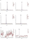
Fig. 2, shows presence of three standard compounds in extract as indicated by retention time. Quantitative analysis was then measured (Table 3) in which HSE contain myricetin (78.23 ng/mg), β-caroten, (0.034 ng/mg) and ascorbic acid (<LOQ). Ascorbic acid was below limit of detection which means that the con- centration of ascorbic acid was very low in HSE. This result was not consistent with previous research that the flower of H. sabdariffa contained β-carotene, phytosterol, citric acid, ascorbic acid, maleic acid, malic acid, niacin, pectin.28
Antioxidants have been studied for their effectiveness in reducing the deleterious effects of aging and agerelated diseases.29 DPPH is very stable free radical and has advantage of being unaffected by side reactions, such as metal ion chelation and enzyme inhibition.30 In the present study, HSE has low DPPH scavenging activity (IC50 = 195.73 µg/mL) compared to ascorbic acid (IC50 = 33.54 µM or 5.91 µg/mL) (Table 4), while IC50 of DPPH activity of myricetin was 4.50 µg/mL as measured in our previous study.19 Myricetin, one of flavonoid compounds, showed highest activitiy among treatments. This is supported by the other study, that flavonoids can scavenge the DPPH radical in a dose-dependent manner, and the DPPH radical scavenging activity (IC50) was decreased in the following order: myricetin (4 µM) > quercetin (5 µM) = luteolin (5µM) > kaempferol (12 µM) > chrysin > apigenin.31 In this study, ascorbic acid also exhibited good scavenging activity, which is validated by other study that ascorbic acid can reduce the generation of reactive oxygen species in some experimental models.3233
HSE showed the lowest ABTS-reducing activity (IC50 = 74.58 µg/mL) compared to myricetin (IC50 = 3.17 µM or 1.01 µg/mL), ascorbic acid (IC50 = 27.38 µM, or 4.82 µg/mL), β-carotene (IC50 = 69.66 µM, or 37.40 µg/mL) (Table 5). These results indicate HSE has weak antioxidant activity compared to three compounds. Myricetin has greater antiradical activity than other flavonoids, and it scavenges oxygen radicals and inhibits lipid peroxidation.34 Myricetin can also act as a free radical scavenger when keratinocytes experience UVB-induced damage.35 Flavonols that contain more hydroxyl groups (one to six OH groups) such as myricetin, have higher free radical and superoxide anion radical scavenging abilities. Myricetin is able to rapidly donate a hydrogen atom to the radicals.31
The present study showed that FRAP activity in concentration-dependent manner, in which higher concentration increased FRAP activity. HSE at the highest concentration (100.00 µg/mL) has moderate activity 46.24 µM Fe(II)/µg, compared to other compounds, myricetin at the highest concentration (3.98 µg/mL) had reduced ferric of 400.81 µM Fe(II)/µg, the highest concentration of β-carotene (6.71 µg/mL) was 10.70 µM Fe(II)/µg, at highest concentration of ascorbic acid (2.10 µg/mL) had value of 288.29 µM Fe(II)/µg (Table 6). HSE has lower antioxidant activity in the FRAP assay compared to myricetin and ascorbic acid. The moderate phenolic and flavonoid content in HSE might be related to its antioxidant activity (Table 1) and contained low myricetin according the UHPLC analysis (Table 3). Another study showed that flavonoids, including myricetin, exhibited antioxidant properties against different types of free radicals.36 Myricetin as flavonoids has greater antiradical activity than other flavonoids, that scavenges oxygen radicals and inhibits lipid peroxidation.37 Myricetin also has effect of antioxidative and collagenase inhibitory activity caused Ultra violet-A (UV-A) in human dermal fibroblasts depends on the number and order of OH groups in those structure compounds.31 The combination of ascorbic acid and vitamin E (α-tocopherol) was reported to synergistically enhance free radical scavenging activity induced by UV in human skin.3839 Ascorbic acid may play a key role in the antioxidant and antiwrinkle effects of the mixture.40
Ultra violet (UV) exposure causes physical changes of the skin through complex pathways and finally generates reactive oxygen species (ROS), matrix metalloproteinases (MMPs) and elastase secretion,41 which require antioxidant to inhibit collagenase and elastase secretion. Collagenase is responsible for extracellular matrix (ECM),42 that inhibit retention of skin elasticity and tensile strength of the skin.43 The present study showed that HSE has low activity in collagenase assay (17.24%) compared to myricetin (99.31%) and was comparable with ascorbic acid (27.10%) and β-carotene (11.42%) at the highest concentration (Fig. 2.A). The most active of collagenase was myricetin with IC50 value 36.60 µM or 11.65 µg/mL, meanwhile the IC50 of ascorbic acid was 465.67 µM or 82.01 µg/mL, HSE was 750.33 µg/mL, and β-carotene was 1,369.50 µM or 734.22 µg/mL (Table 7). The result of present study showed moderate collagenase inhibitory activity of HSE compared to other compounds. However, myricetin exhibited the highest collagenase inhibitory activity. As reported in previous study, myricetin inhibited the wrinkle formation formation in UVB irradiation in mouse skin, that might block damage to the basement membranes by inhibiting expression and activity of MMP-9.44
Elastin, an ECM protein, is most abundant in organs that provides elasticity to the connective tissues. Therefore, inhibition of the elastase activity ingredients could be used in cosmetic formulation to protect against skin aging and wrinkles.8 Myricetin exhibited the most active with the highest elastase inhibitory activity 92.86% and IC50 value 1.20 µg/mL or 3.76 µM. HSE exhibited the highest elastase inhibitory activity IC50 value 103.83 µg/mL, meanwhile the IC50 value of ascorbic acid was 15.47 µg/mL or 87.83 µM and the IC50 value of β-carotene was 79.95 µg/mL or 148.92 µM and (Fig. 2B, Table 7). These findings were supported with previous study of flavonoid effects on elastase release at the final concentration range of 0.5 to 10 µmol/L. Myricetin inhibited elastase release at the highest concentration tested (10 µmol/L).45
Hyaluronidase inhibitory was determined by measuring the amount of N-acetylglucosamine splited from sodium hyaluronate.46 HSE has highest IC50 value (619.43 µg/mL) compared to ascorbic acid (160.58 µM or 28.28 µg/mL), β-carotene (34.21 µM or 18.35 µg/mL) and myricetin (21.30 µM or 6.78 µg/mL). Myricetin has highest antiaging activity of all treatments. Myricetin might strongly block damage in basement membranes that can prevent UVBinduced wrinkle formation. β-carotene also plays an important role in the aging process as a mediator in redox processes.47 It has been found that ascorbic acid and β-carotene are potent as radiation protectors.48 Ascorbic acid and β-carotene can act as antiaging compounds and protect cell membrane from oxidative stress.47 In vivo study in pig skin demonstrated that the combination of topical ascorbic acid and vitamin E synergistically provide protection against UV-induced erythema and formation of sunburn cells and thymine dimers.4950 Ascorbic acid, vitamin E and polyphenols were reported to have significant antiaging effects.51 Those compounds have potency to inhibit hyaluronidase by lowering its viscosity and increasing the permeability.52

Figures and Tables

Fig. 1

Chemical structure of compounds in the H. sabdariffa. (1) Myricetin (C15H10O8), (2) Ascorbic acid (C6H8O6), (3) β-carotene (C40H56).

nps-23-192-g001
Fig. 2

Chromatogram of UHPLC Analysis. (1) HSE and myricetin, (2) HSE and β-carotene, (3) HSE and ascorbic acid.

nps-23-192-g002
Fig. 3

Effect of HSE (µg/mL), myricetin, β-carotene, ascorbic acid (µM) on collagenase, elastase, and hyaluronidase inhibitory activity. (1) Collagenase inhibitory activity, (2) Elastase inhibitory activity, (3) Hyaluronidase inhibitory activity.

nps-23-192-g003
Table 1

The result of qualitative phytochemical screening of HSE (phenols, steroids/triterpenoids, terpenoids, saponins, flavonoids, tannins and alkaloids)

nps-23-192-i001
Phytochemical content HSE
Phenols ++
Steroids/Triterpenoids −/+
Terpenoids
Saponins
Flavonoids ++
Tanins ++
Alkaloids +

++++: very high content; +++ : high content; ++ : moderate content; + : low content; − : not detected

Table 2

Total phenolic content of HSE with gallic acid standard.

nps-23-192-i002
Sample Linear Equation R2 Total phenolic content (µg GAE/mg)
HSE y = 0.003x + 0.024 0.998 23.85 ± 1.17

The phenolic content of HSE was measured in triplicate.

In triplicate. Linear equation (Y = 0.003x + 0.024), coefficient of regression (R2 = 0.998) of Gallic Acid was calculated. The data was presented as mean ± standard deviation.

Table 3

UHPLC analysis of compounds contained in HSE, quantification of HSE was performed with LC-MS/MS using myricetin, β-carotene, and ascorbic acid as standard. The UHPLC analysis were performed in duplicate.

nps-23-192-i003
No Compounds Weight (g) Area Measured-Content (µg/mL) Calculated-Content (µg/mL)
1 Myricetin 1.40 1,027 21.91 78.23
2 β-carotene 1.80 8,817 0.361 0.034
3 Ascorbic Acid 1.40 0.01 < LOQ
Table 4

DPPH scavenging activity of HSE, myricetin, β-carotene, and ascorbic acid.

nps-23-192-i004
Samples The highest activity of ABTS reducing activity (%) IC50 (µg/mL) IC50 (µM)
HSE 76.79 195.73 ± 18.63
β-carotene 48.65 222.95 ± 9.62 415.27 ± 17.91
Ascorbic Acid 95.94 555.91 ± 0.66 533.54 ± 3.77

The data was presented as median inhibition concentration (IC50). The DPPH scavenging activity were measured in triplicate for each concentration of samples. Each DPPH scavenging activity of samples were calculated the IC50 and each sample had three value of IC50. The IC50 value were presented as mean ± standard deviation, the IC50 value were presented only µg/mL for HSE, both µg/mL and µM for β-carotene, ascorbic acid.

Table 5

ABTS-reducing activity of HSE, myricetin, β-carotene, and ascorbic acid were presented as IC50.

nps-23-192-i005
Samples The highest activity of ABTS reducing activity (%) IC50 (µg/mL) IC50 (µM)
HSE 34.37 74.58 ± 2.97
Myricetin 75.66 1.01 ± 0.02 3.17 ± 0.06
β-carotene 37.30 37.40 ± 6.69 69.66 ± 12.47
Ascorbic Acid 88.33 4.82 ± 1.19 27.38 ± 6.76

The ABTS-reducing activity were measured in triplicate for each concentration of samples. Each ABTS-reducing activity of samples were calculated the IC50 and each sample had three value of IC50. The IC50 value were presented as mean ± standard deviation.

Table 6

FRAP activity of HSE, myricetin, β-carotene, and ascorbic acid.

nps-23-192-i006
Concentrations HSE (µg/mL) HSE FRAP Activity (µM Fe(II)/µg) Concetrations Myricetin (µg/mL) Myricetin FRAP Activity (µM Fe(II)/µg) Concetrations β-carotene (µg/mL) β-carotene FRAP Activity (µM Fe(II)/µg) Concetrations Ascorbic acid (µg/mL) Ascorbic Acid FRAP Activity (µM Fe(II)/µg)
100.00 46.24 ± 0.13a 3.98 400.81 ± 4.10a 6.71 10.70 ± 0.24a 2.10 288.29 ± 9.49a
50.00 21.84 ± 0.60b 1.99 328.53 ± 4.19b 3.36 4.27 ± 0.03b 1.10 139.52 ± 5.99b
25.00 8.61 ± 0.20c 0.99 169.77 ± 17.72c 1.68 1.07 ± 0.13c 0.55 65.44 ± 1.83c
12.50 2.66 ± 0.34d 0.50 87.86 ± 11.40d 0.84 −1.28 ± 0.64d 0.28 32.25 ± 1.84d

The data was presented as mean ± standard deviation. The FRAP activity were measured in triplicate for each concentration of samples. The concentrations level of samples were expressed µg/mL for HSE, µg/mL and µM for compounds (myricetin, β-carotene, ascorbic acid). Results of FRAP activity were expressed in µM Fe(II)/µg sample. Different superscripts in the same column (a, b, c, d) indicate significant differences among the means of groups (concentrations of HSE, myricetin, β-carotene, ascorbic acid) based on Tukey's HSD Posthoc comparisons (P < 0.05).

Table 7

The IC50 value of collagenase, elastase, and hyaluronidase inhibitory activity of HSE, myricetin, β-carotene and ascorbic acid.

nps-23-192-i007
Samples IC50
Collagenase inhibitory (µg/mL) Collagenase inhibitory (µM) Elastase inhibitory (µg/mL) Elastase inhibitory (µM) Hyaluronidase inhibitory (µg/mL) Hyaluronidase inhibitory (µM)
HSE 750.33 ± 37.51 103.83 ± 4.00 619.43 ± 42.02
Myricetin 11.65 ± 2.08 36.60 ± 6.53 1.20 ± 0.11 3.76 ± 0.34 6.78 ± 0.75 21.30 ± 2.37
β-carotene 735.24 ± 102.29 1369.50 ± 190.53 79.95 ± 1.85 148.92 ± 3.44 18.35 ± 3.65 34.21 ± 6.80
Ascorbic Acid 82.01 ± 4.29 465.67 ± 24.37 15.47 ± 0.24 87.83 ± 1.38 28.28 ± 3.04 160.58 ± 17.27

The IC50 was presented as mean ± standard deviation, it was only µg/mL for HSE, both µg/mL and µM for β-carotene, ascorbic acid. The antiaging activity (collagenase, elastase, hyaluronidase inhibitory) were measured in triplicate for each concentration of samples.

Acknowledgements

This research was supported by Biomolecular and Biomedical Research Center, Aretha Medika Utama, Bandung, Indonesia for research grant, laboratory facilities and research methodology.

References

1. Benaiges A, Marcet P, Armengol R, Betes C, Gironés E. Int J Cosmet Sci. 1998; 20:223–233.
2. Jenkins G. Mech Ageing Dev. 2002; 123:801–810.
3. Schlotmann K, Kaeten M, Black AF, Damour O, Waldmann-Laue M, Förster T. Int J Cosmet Sci. 2001; 23:309–318.
4. McCullough JL, Kelly KM. Ann N Y Acad Sci. 2006; 1067:323–331.
5. Aslam MN, Lansky EP, Varani J. J Ethnopharmacol. 2006; 103:311–318.
6. Chanchal D, Swarnlata S. J Cosmet Dermatol. 2008; 7:89–95.
7. Farris PK. Cosmetic Dermatol. 2003; 16:59–72.
8. Roy A, Sahu RK, Matlam M, Deshmukh VK, Dwivedi J, Jha AK. Pharmacogn Rev. 2013; 7:97–106.
9. Pandel R, Poljšak B, Godic A, Dahmane R. ISRN Dermatol. 2013; 2013:930164.
10. Taniguchi M, Arai N, Kohno K, Ushio S, Fukuda S. Eur J Pharmacol. 2012; 674:126–131.
11. Johnston MF, Hili P, Naughton DP. BMC Complement Altern Med. 2009; 9:1–11.
12. Tsai PJ, McIntosh J, Pearce P, Camden B, Jordan B. Food Res Int. 2002; 35:351–356.
13. Mohamad O, Mohd B, Abdul M, Herman S. Bull PGM. 2002.
14. Widowati W, Wijaya L, Wargasetia TL, Bachtiar I, Yellianty Y, Laksmitawati DR. J Exp Integr Med. 2013; 3:225–230.
15. Widowati W, Ratnawati H, Rusdi U, Winarno W, Imanuel V. HAYATI J Biosci. 2010; 17:85–90.
crossref
16. Bera TK, Chatterjee K, Ghosh D. Int J Ayurveda Res. 2010; 1:18–24.
17. Adnyana IK, Abuzaid AS, Iskandar EY, Kurniati NF. . Int J Med Res Health Sci. 2016; 5:23–28.
18. Widowati W, Ratnawati H, Husin W, Maesaroh M. Biomed Eng. 2015; 1:24–29.
19. Widowati W, Herlina T, Ratnawati H, Constantia G, Deva I, Maesaroh M. Biol Med Nat Prod Chem. 2015; 4:35–39.
20. Lucini L, Pellizzoni M, Baffi C, Molinari GP. J Sci Food Agric. 2012; 92:1297–1303.
21. Sohn DH, Kim YC, Oh SH, Park EJ, Li X, Lee BH. Phytomedicine. 2003; 10:165–169.
22. Dasgupta A, Ray D, Chatterjee A, Roy A, Acharya K. J Pharm Biol Chem Sci. 2014; 5:510–520.
23. Widowati W, Widyanto RM, Husin W, Ratnawati H, Laksmitawati DR, Setiawan B, Nugrahenny D, Bachtiar I. Iran J Basic Med Sci. 2014; 17:702–709.
24. Mishra A, Bapat MM, Tilak JC, Devasagayam TT. Curr Sci. 2006; 91:90–93.
25. Widowati W, Fauziah N, Herdiman H, Afni M, Afifah E, Kusuma HSW, Nufus H, Arumwardana S, Rihibiha DD. J Nat Rem. 2016; 16:89–99.
26. Tu PT, Tawata S. Molecules. 2015; 20:16723–16740.
27. Bergmeier D, Berres PHD, Filippi D, Bilibio D, Bettiol VR, Priamo WL. Maringa. 2014; 36:545–551.
28. Mahadevan N, Shivali , Kamboj P. Nat Prod Radiance. 2009; 8:77–83.
29. Fusco D, Colloca G, Lo Monaco MR, Cesari M. Clin Interv Aging. 2007; 2:377–387.
30. Amarowicz R, Pegg RB, Rahimi-Moghaddam P, Barl B, Weil JA. Food Chem. 2004; 84:551–562.
31. Oliveira CP, Kassab P, Lopasso FP, Souza HP, Janiszewski M, Laurindo FR, Iriya K, Laudanna AA. World J Gastroenterol. 2003; 9:446–448.
32. Wang X, Falcone T, Attaran M, Goldberg JM, Agarwal A, Sharma RK. Fertil Steril. 2002; 78:1272–1277.
33. Huang JH, Huang CC, Fang JY, Yang C, Chan CM, Wu NL, Kang SW, Hung CF. Toxicol In Vitro. 2010; 24:21–28.
34. Formica JV, Regelson W. Food Chem Toxicol. 1995; 33:1061–1080.
35. Sim GS, Lee BC, Cho HS, Lee JW, Kim JH, Lee DH, Kim JH, Pyo HB, Moon DC, Oh KW, Yun YP, Hong JT. Arch Pharm Res. 2007; 30:290–298.
36. Wang L, Tu YC, Lian TW, Hung JT, Yen JH, Wu MJ. J Agric Food Chem. 2006; 54:9798–9804.
37. Mattivi F, Guzzon R, Vrhovsek U, Stefanini M, Velasco R. J Agric Food Chem. 2006; 54:7692–7702.
38. Swindells K, Rhodes L. Photodermatol Photoimmunol Photomed. 2004; 20:297–304.
39. Boelsma E, Hendriks H, Roza L. Am J Clin Nutr. 2001; 73:853–864.
40. Haftek M, Mac-Mary S, Le Bitoux MA, Creidi P, Seité S, Rougier A, Humbert P. Exp Dermatol. 2008; 17:946–952.
41. Demeule M, Brossard M, Pagé M, Gingras D, Béliveau R. Biochim Biophys Acta. 2000; 1478:51–60.
42. Pientaweeratch S, Panapisal V, Tansirikongkol A. Pharm Biol. 2016; 54:1865–1872.
43. Roy A, Sahu RK, Matlam M, Deshmukh VK, Dwivedi J, Jha AK. Pharmacogn Rev. 2013; 7:97–106.
44. Jung SK, Lee KW, Kim HY, Oh MH, Byun S, Lim SH, Heo YS, Kang NJ, Bode AM, Dong Z, Lee HJ. Biochem Pharmacol. 2010; 79:1455–1461.
45. Kanashiro A, Souza JG, Kabeya LM, Azzolini AE, Lucisano-Valim YM. Z Naturforsch C. 2007; 62:357–361.
46. Sahasrabudhe A, Deodhar M. Int J Bot. 2010; 6:299–303.
47. Getoff N. Rad Phys Chem. 2007; 76:1577–1586.
48. Kammerer C, Czermak I, Getoff N. Radiat Phys Chem. 2001; 60:71–72.
49. Darr D, Dunston S, Faust H, Pinnell S. Acta Derm Venereol. 1996; 76:264–268.
50. Lin JY, Selim MA, Shea CR, Grichnik JM, Omar MM, Monteiro-Riviere NA, Pinnell SR. J Am Acad Dermatol. 2003; 48:866–874.
51. Rizvi S, Jha R. Expert Opin Drug Discov. 2011; 6:89–102.
52. Girish KS, Kemparaju K. Life Sci. 2007; 80:1921–1943.
TOOLS
Similar articles